
Page 113 - Haematologica-5
P. 113
Ruxolitinib in advanced relapsed/refractory HL
baseline and after the first cycle of treatment. At baseline, there was no difference in cytokine levels between responders and non-responders. In responders, the only cytokine that decreased significantly was CX-CL10 (P=0.01). In patients presenting with pruritus (n=11), the levels of platelet-derived growth factor-BB (PDGF-BB) (Online Supplementary Appendix), interleukin (IL)-5, IL-10, IL-12, IL-13, IL-17, eotaxin, fibroblast growth factor basic (FGF basic), macrophage inflammatory protein 1b (MIP1b), regulated on activation, normal T-cell expressed and secreted (RANTES), and vascular endothelial growth factor (VEGF) were significantly increased. In the latter patients, ruxolitinib treatment significantly decreased the levels of PDGF-BB, IL-10, IL-12, IL-13, IL-17, FGF basic and VEGF. Among the patients who could be analyzed for JAK2 amplification in HRS cells (n=12), polysomy (sug- gesting hyperdiploidy) was detected in all of them, and specific JAK2 amplification in only one. This latter patient
achieved a partial response as determined by computed tomography criteria and also a positron emission tomog- raphy-determined response lasting 4 months. It is note- worthy that the PDL1 and PDL2 loci (which are in the vicinity of the JAK2 locus at 9p24), analyzed by fluores- cent in situ hybridization with bacterial artificial chromo- some probes, showed the same pattern of gains as for the JAK2 locus.
Discussion
JAK/STAT activation, driven by an aberrant network of cytokines and chemokines in the HL microenvironment, is critical for the proliferation and survival of neoplastic HRS cells.24,25 The JAK/STAT pathway also plays a role in immune evasion by HL cells via the secretion of chemokines leading to Th2 homing or via the regulation
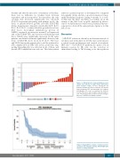
Figure 4. Waterfall plot demonstrating percent change from baseline in target tumor dimensions (best response, n=27). Of note, among the 32 patients evaluable for disease response, five had no end-of-treatment SPD measurements by computer tomography (CT) scan as planned by protocol because there were obvious signs of disease progres- sion. *Persisting positive positron emission tomogra- phy scan, considered as partial response.
Figure 5. Kaplan-Meier estimate of progression-free survival in 32 evaluable patients with Hodgkin lym- phoma receiving ruxolitinib.
haematologica | 2018; 103(5)
845
