
Page 110 - Haematologica-5
P. 110
E. Van Den Neste et al.
Study assessments
Baseline assessments comprised documentation of disease- related symptoms, physical examination, laboratory tests, and imaging studies of the neck, chest, abdomen, and pelvis, using computed tomography or magnetic resonance imaging. Biopsy prior to inclusion was recommended, but not mandatory. Tumors were measured at baseline, at the end of every two cycles of rux- olitinib, and following the six-cycle induction, as well as during maintenance therapy. Given the exploratory nature of the study, there was no centralized review of computed tomography response. However, positron emission tomographic images of the responders were all centrally reviewed by a nuclear physician (ASC) to confirm partial or complete metabolic response based on the Deauville five-point scale. The evaluable study population for efficacy was restricted to patients who had received at least 28 days of the study drug.
Safety was monitored for up to 1 month after treatment. Adverse events were summarized by means of the Medical Dictionary for Regulatory Activities, and graded using the National Cancer Institute’s Common Terminology Criteria for Adverse Events (NCI-CTCAE), version 3.0. Laboratory abnormal- ities were assessed according to NCI-CTCAE version 4.0. Only grade 3 or 4 toxicities and grade 2 infections were to be reported. All patients were included in the toxicity analysis.
Exploratory biomarker analysis
Blood samples (5 mL) were taken at baseline prior to drug administration and on day 1 of cycle 2 for the measurement of 27 cytokines related to the immune system using bead-based immunoassays. JAK2 gains, amplifications, and gene rearrange- ments were also investigated using fluorescent in situ hybridiza- tion with two tri-color sets of probes associating JAK2/9p24 break-apart probes with a control centromeric probe (CEP9/9q21): the already prepared probes from Empire genomics on the one hand, and the association of the JAK2 B/A probe from Kreatech with the CEP9 probe from Vysis on the other hand. The CD274/PDL1 and PDCD1LG2/PDL2 loci at 9p24 were studied with home-made prepared bacterial artificial chromosome probes purchased from the Chori BACPAC Resources Center (Oakland, CA, USA). Extraction, labeling and hybridization were performed on paraffin-embedded tissue, as previously reported.22
Statistical methods
The sample size for this phase II study was calculated using an exact single-stage phase II design.23 A two-stage design with interim analysis for activity or toxicity was not planned given the very advanced stage of the patients, the relative paucity of alternative options, and the potential toxicity of ruxolitinib that was expected to be in the low range, based on myelofibrosis data. The treatment was considered ineffective if the ORR was ≤15%, and effective if the ORR was ≥35%. Under the assump- tion of an alpha first-order risk error set at 5% and beta at 20% with a one-sided test, it was deemed necessary to include a total of 28 evaluable patients with a cut-off number of eight. If at least eight patients had a response, the hypothesis of an ORR ≤15% was rejected with both a target error rate and an actual error rate of 0.05. If seven or fewer patients had a response, the hypothesis of an ORR ≥35% was rejected with a target error rate of 0.2 and an actual error rate of 0.187. The ORR estimate and its 90% confidence intervals (CI) were calculated for all patients who completed at least one cycle of the study drug.
The Kaplan-Meier method was employed to estimate the median value and its 95% CI for time to response, duration of response, progression-free survival and overall survival. The
safety analysis comprised all patients who received at least one dose of the study drug. All statistical analyses were performed using SAS software, version 9.2. P-values <0.05 were consid- ered statistically significant. All available data were included in data listings and tabulations, with no imputations of values for missing data. An interim analysis was neither planned nor per- formed.
Results
Patients’ disposition and characteristics
The patients’ characteristics are listed in Table 1. From July 2013 to December 2014, a total of 33 patients with R/R HL were recruited. Their median age was 37 years (range, 19-80). Most of the patients had advanced HL (stage III/IV) and had been heavily pretreated, with a median number of five prior regimens including autolo- gous SCT (54%), allogeneic SCT(15%), and BV (82%). Of the 33 patients recruited, 27 (82%) had refractory HL and 22 had biopsy-confirmed relapse of HL. Among the six patients displaying a response, a biopsy was per- formed in five of them at relapse [8 days, 12 days, 6 weeks (n=2) and 14 months prior to inclusion in the study].
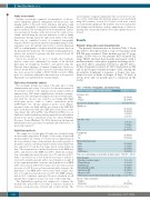
Table 1. Patient’s demographics and characteristics.
Patients’ demographics and characteristics
Gender, n (%) Male
Female
Age in years, median (range)
ECOG score 0
1 2 3
Ann Arbor stage I
II III IV
B symptoms Yes
No
Extranodal involvement Bone
Liver
Lung
Soft tissues
Time since initial diagnosis in months, median (range)
Prior therapies
Prior lines, median (range)
Chemotherapy
Radiotherapy
Brentuximab vedotin
Autologous SCT
Allogeneic SCT Intervalsincelasttreatmentinmonths,median(range) 6(1.1–75.0)
Disease status at inclusion
Relapse 6 Refractory 27 (81.8%)
SCT: stem cell transplantation; ECOG: Eastern Cooperative Oncology Group.
All patients (n=33)
21 (63.6%) 12 (36.4%)
37.0 (19.0-80.0)
11 (33.3%) 15 (45.5%) 5 (15.2%) 2 (6.1%)
1 (3.0%) 7 (21.2%) 3 (9.1%) 22 (66.7%)
16 (48.5%) 17 (51.5%)
13 (39.4%) 6 (18.2%) 12 (36.4%) 4 (12.1%)
55.4 (8.7 – 216.1)
5 (1 – 16) 33 (100%) 18 (54.5%) 27 (82%) 18 (54.5%) 5 (15.2%)
842
haematologica | 2018; 103(5)
