
Page 72 - Haematologica-April 2018
P. 72
616
A. Thivakaran et al.
between GFI1B expression level and SPI1 expression as well as MEIS1 and a positive correlation with MLLT3 (Online Supplementary Figure S1).
Reduced expression level or loss of Gfi1b promotes progression of MDS to AML in a murine MDS model
To investigate a connection between Gfi1b level and AML, we used different mouse strains and models of human leukemia. We used one strain in which both Gfi1b alleles can be conditionally deleted in the hematopoietic system upon poly(I:C) administration, resembling absence of Gfi1b expression (Gfi1bfl/flMxCretg).6 In a second mouse model, one coding allele of Gfi1b is replaced by enhanced green fluorescence protein (EGFP) cDNA (Gfi1bEGFP/wt),13 which leads to a lower expression level of Gfi1b (see below). Finally, wild-type mice were used to model normal/high Gfi1b expression. To study whether reduced Gfi1b expression accelerates MDS to AML pro- gression, we crossed the above-mentioned mouse strains with NUP98/HOXD13tg mice, which represent a model for human MDS/AML.34
We first used the Gfi1b:EGFP knock-in reporter mouse strain and crossed these mice with NUP98/HOXD13tg mice (Figure 3A). Loss of one allele of Gfi1b shortened the latency period of AML development (Figure 3B). In BM cells derived from heterozygous leukemic mice, the expression of Gfi1b mRNA and Gfi1b protein levels were reduced to approximately 50% compared to BM cells from Gfi1bwt/wt leukemic mice (Online Supplementary Figure S2A and B). Furthermore, we found that the EGFP expres- sion level and hence Gfi1b expression level was signifi- cantly lower in the myeloid blasts when the disease onset was within the first 250 days compared to Gfi1b expres- sion in blasts from mice that developed overt leukemia more than 250 days after birth (Figure 3C). The leukemic cells from Gfi1bwt/wt or Gfi1bEGFP/wt animals showed no sig- nificant differences with regard to surface marker expres- sion, spleen size, white blood cell and platelet counts, or cytological appearance, but showed significant differ- ences with regard to hemoglobin and red blood cells (Figure 3D, Online Supplementary Figure S2C-F and data not shown), which might be due to a potential dose-depen- dent role of Gfi1b in erythropoiesis.6,9
We next examined how complete absence of Gfi1b influences MDS to AML progression. We used the Gfi1b conditional knockout mouse model (Gfi1bfl/flMxCretg), whereby the expression of Gfi1b can be conditionally abrogated in the hematopoietic system upon poly(I:C) administration6 (Figure 3E). The absence of Gfi1b resulted in a substantially earlier onset of AML with a median sur- vival time of approximately 50 days (P<0.0001) (Figure 3F). Cre-mediated excision was verified to be efficient in leukemic Gfi1bfl/flMxCretgNUP98/HOXD13tg mice after poly(I:C) administration with non-excised Gfi1b alleles below detection levels (Figure 3G), and this was associat- ed with practically no expression of Gfi1b mRNA and protein (Online Supplementary Figure S2A and B).6 Leukemic cells from Gfi1bfl/flMxCretg and Gfi1bfl/flMxCretg animals showed no significant differences in spleen size, white blood cells or cytological appearance but significant differences in hemoglobin, red blood cells and platelet counts (Figure 3H and Online Supplementary Figure S2G-J and data not shown), which might be due to a dose-depen- dent role of Gfi1b in erythropoiesis.6,9 The absence of Gfi1b led to a reduced frequency of myeloid cells (Figure 3I, left, middle, and Online Supplementary Figure S3A-C). CD117 (c-Kit) was uniformly higher expressed on all Gfi1b-deficient blast cells (derived from Gfi1bfl/flMxCretg) mice compared to Gfi1b expressing blasts (Gfi1bfl/flMxCrewt) (Figure 3I, right). Finally, there was no difference with regard to apoptosis in NUP98/HOXD13tg mice (Online Supplementary Figure S3D). In our murine model of MDS/AML development, we did not observe a positive correlation between Gfi1b and Mllt3 expression nor a negative correlation between Gfi1b and Spi1 expres- sion, which might be disease context-dependent and thus not reproducible in all types of AML (Online Supplementary Figure S3A and B). We also analyzed the expression level of Meis1, since Chowdhury et al. observed a negative correlation between GFI1B and MEIS1.33 We were able to confirm this finding for this model of AML (Online Supplementary Figure S4C).
Loss of Gfi1b promotes the progression of myelopro- liferative disorder in a conditional Kras mouse model
To validate the results above in a second model, we used mice conditionally expressing a mutated form of
ABC
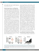
Figure 1. Correlation between different GFI1B expression levels and myelodysplastic syndrome (MDS) or acute myeloid leukemia (AML) prognosis. (A) Expression of GFI1B in CD34+ AML cells (n=269) compared to CD34+ control cells (n=8) based on the patient cohort published by Valk et al.;21 P≤0.0001. (B) Expression of GFI1B in CD34+ MDS cells (n=23) compared to CD34+ AML cells (n=501) based on the patient cohort published by Wouters et al.;19 P≤0.0001. (C) Expression of GFI1B in leukemic stem cells (LSCs)20 of different AML subtypes compared to normal hematopoietic stem cells (HSCs) or common myeloid progenitor cells (CMPs) in published gene expression arrays.31
haematologica | 2018; 103(4)
