
Page 60 - Haematologica3
P. 60
420
R. Bottega et al.
site-directed mutagenesis using specific primers (available upon request) and transfected, using calcium phosphate, in 293T cells treated with 2 mM hydroxyurea (HU) for 24 hours.17 Protein whole and fractionated cell extracts were prepared using M-PERTM Mammalian Protein Extraction Reagent and NE-PERTM Nuclear and Cytoplasmic Extraction Reagents (Thermo Fisher Scientific), respectively. Primary antibodies were used as follows: anti- FANCA (Savino et al.,18 1:500), anti-FANCD2 (Santa Cruz, sc- 20022, 1:500), add anti-HSP90 (Santa Cruz, sc-7947, 1:4000), anti- ORC2 (Abcam, ab68348; 1:500), anti-FLAG (OctA Probe H5, Santa Cruz, 1:500), and anti-α Tubulin (Santa Cruz, sc-5286, 1:2000). Immuno-reactivity was visualized using the Enhanced Chemiluminescent SuperSignalTm West Femto Maximum Sensitivity Substrate (Pierce).
For immunofluorescence assay, anti-FLAG antibody was used
with anti-mouse FITC secondary antibody (F0479, DakoCytomation), while nuclei were stained with 1 ug/mL pro- pidium iodide solution (Sigma Aldrich).
Biochemical assays
FANCA pcDNA3.1 constructs were transfected in LFB cells not expressing the FANCA protein (FANCA-/-) using Lipofectamine (Thermo Fisher Scientific). Biochemical assays (ATP/AMP evalua- tion, electron transfer between complex I to complex III and Oxygen consumption measurements) were performed as described in Columbaro et al.19
Fo-F1 ATP synthase activity assay
Evaluation of the Fo-F1 ATP synthase activity was performed as previously described.20 Briefly, 200,000 cells were incubated for 10
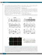
A
B
C
DE
Figure 2. Expression of p.His913Pro and p.Arg951Gln FANCA proteins. (A) Western blot of total lysates from lymphoblast (LFB) cells of patients carrying the p.His913Pro (F3, F4, F5 and F6) and the p.Arg951Gln (F7 and F10) mutations, showing that the mutant FANCA proteins are expressed. The controls are wild-type (+/+) LFB cells and LFB cells not expressing the FANCA protein (-/-) due to a homozygous large intragenic deletion (c.284-?_1826+?del) of FANCA. (B) Western blot of cytoplasmic (C) and nuclear (N) fractionated cellular lysates after 2 mM hydroxyurea treatment (24 hours) of LFB from patients F3 and F7, showing that the endogenous p.His913Pro and p.Arg951Gln proteins do not translocate to the nucleus. Controls +/+ and -/-, as indicated in (A). (C) Western blot of cytoplasmic (C), nuclear (N) fractionated, or total (TL) cellular lysates from 293T cells transfected with the wild-type (wt) or the mutant (H913P and R951Q) forms of FANCA tagged with FLAG, confirming the exogenous p.His913Pro and p.Arg951Gln FANCA proteins are retained in the cytoplasm. (D) Immunofluorescence analyses on 293T cells transfected as indicated in (C). Nuclei are stained with propidium iodide (PI). (E) Western blot of different LFB cells exposed to 2 mM hydroxyurea (24 hours) showing no monoubiquitination of the FANCD2 protein. Control +/+, as indicated in (A).
haematologica | 2018; 103(3)
