
Page 51 - Haematologica3
P. 51
miR-451 inhibits Cab39 for stress erythropoiesis
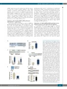
mRNA expression via direct interaction with this region, we fused the 3′-UTR of Cab39 mRNA to the coding sequence of luciferase cDNA (Figure 4F). In 293T cells, luciferase reporter activity was inhibited approximately 200-fold after co-expression of miR-451. Mutations in the Cab39 mRNA 3’ UTR that disrupt complementarity to the miR-451 seed sequence abrogated repression of reporter activity. Together, these findings verify that miR-451 inhibits Cab39 mRNA expression directly and that this interaction occurs during erythropoiesis.
Activation of the Cab39/AMPK/mTOR pathway in miR-144/451−/− erythroblasts
Cab39 binds LKB1 and STRAD to activate AMPK by phosphorylating the protein at Thr172 (Figure 5A).22,23 Of note, LKB1 and AMPK mRNAs are up-regulated during normal mouse and human erythroid maturation (Online Supplementary Figure S2A-E).19,24 Cab39 was up-regulated in cultured miR-144/451−/− FL erythroblasts compared to con- trols (Figures 4D and 5B and C). These cells also exhibited strongly increased phosphorylation of AMPK at Thr172, and to a lesser extent, upregulation of total AMPK protein (Figure 5B and D). AMPK can inhibit the mTOR pathway to inhibit cell growth and either induce or suppress apop- tosis, depending on cellular context.25 To investigate this
AB
C
D
mechanism in miR-144/451−/− erythroblasts, we performed Western blot studies to interrogate upstream and down- stream effectors.26,27 miR-144/451−/− FL erythroblasts grown in culture exhibited elevated phospho-Raptor and phos- pho-TSC2, along with reduced phosphorylation of p70S6K, S6, and eIF4B (Figure 5B and E-I), consistent with suppression of the mTOR by activated AMPK (Figure 5A). We observed similar patterns in primary erythroblasts from E14.5 FLs (Online Supplementary Figure S3). Together, these findings indicate that the loss of miR-144/451 during FL erythropoiesis derepresses the miR-451 target Cab39, resulting in mTOR repression.
Attenuation of Cab39/AMPK/mTOR signaling rescues erythroid apoptosis after miR-144/451 depletion
To determine whether miR-144/451 regulates survival of FL erythroblasts via Cab39/AMPK/mTOR, we designed shRNA-expressing retroviruses to knock down compo- nents of this pathway. We grew FL erythroblasts in expan- sion medium, infected them with individual shRNA retro- viruses, induced erythroid maturation for 24 h, and then examined apoptosis in the cells. Inhibiting Cab39 expres- sion in miR-144/451−/− erythroblasts by approximately 55- 70% with 2 different shRNAs reduced apoptosis by approximately 45% (Figure 6A-C). In contrast, no signifi-
Figure 4. miR-451 targets Cab39 mRNA in erythrob- lasts. (A) Nucleotide sequence alignments showing partial complementarity between the mouse and human 3′-UTRs of Cab39 mRNAs and miR-451. The miR-451 seed-sequence recognition sites in the mRNAs are boxed in white. (B) Fold change in the Cab39 mRNA level from quantitative RT-PCR analy- sis. Ter119+ nucleated cells were sorted from bone marrow, and a microarray was performed using an Affymetrix GeneChip.15 N=3 mice were used. **P<0.01 (t-test). (C) Cab39 protein levels in both spleen and bone marrow miR-144/451−/− erythrob- lasts were increased relative to those in wild-type (+/+, WT) controls (top). Ter119+ cells were purified from bone marrow, and whole-cell lysates were analyzed via Western blots with the Cab39 antibody. Representative results for 2 mice of each genotype are shown. (Bottom) Quantitative image analysis from multiple experiments. (D) Cab39 protein levels in miR-144/451−/− fetal liver (FL) erythroblasts were increased relative to those in WT controls (top). (Bottom) Quantitative image analysis for 3 WT and 6 miR-144/451−/− (KO) FLs. (E) G1E proerythroblast cells were transduced with retrovirus encoding miR- 451 or empty vector as control. Transduced cells were selected with puromycin and analyzed for Cab39 via Western blots. (Top) Results of a represen- tative experiment and (Bottom) of a quantitative analysis of the Western blot signal intensity from 3 independent experiments. **P<0.01 (t-test). (F) Interaction between miR-451 and the Cab39 3′-UTR inhibits the expression of a linked reporter gene. Firefly luciferase cDNA was fused to the normal 3′- UTR of Cab39 cDNA or a mutant version (mt) contain- ing a 3-bp mutation within the region complementary to the miR-451 seed sequence. The reporter constructs were cloned into an expression vector and transfect- ed into 293T cells, along with an miR-451 expression construct and a constitutively active Renilla luciferase control plasmid. Luciferase activities were determined 24 hours post transfection. (Bottom) Bars represent the Firefly/Renilla luciferase activity; levels from the reporter vector lacking the Cab39 3′- UTR were assigned an arbitrary value of 1. Results are given as the average of 3 separate experiments. **P<0.01 (t-test). Hsa: human; Mmu: mouse.
EF
haematologica | 2018; 103(3)
411
