
Page 188 - Haematologica3
P. 188
M. Nagy et al.
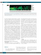
Figure 6. Altered thrombus formation after bone marrow transplantation. Thrombus formation (1600 s-1) on three microspots measured with blood from indicated home controls (HC1-12), travel controls (C1-6) and 2 patients P1 (ORAI1W/W) and P7 (FERMT3R/X), both before (*) and after bone marrow transplantation (BMT). For description of microspots and parameters, see Figure 4. Subtraction heatmap of scaled values after filtering for differences considered to be relevant, i.e. outside the range of mean ± 2 SD (HC1-12).
548
late with low thrombus formation parameters, especially on spot 1. By comparison with flow studies using blood from healthy controls at lower counts, it appears that mild thrombocytopenia can lead to a more severe reduction in thrombus formation if combined with lower platelet func- tionality. Mild thrombocytopenia for patients with a loss- of-function mutation in ORAI1 or STIM1 has been report- ed before.16 So far, published papers on patients with a FERMT3 mutation have not reported on thrombocytope- nia.26,27
Because of the relatively large number of patients in our study, we were able to separate the thrombus formation parameters linked to qualitative or quantitative platelet defects. Both unsupervised clustering of the heatmap data and PCA of the raw data indicated that mostly thrombus parameters on spot 1 (collagen; platelet receptors GPIb-V- IX, GPVI, α2β1) showed a dependency on platelet count, whereas those of spot 2 (VWF/rhodocytin; receptors GPIb-V-IX, CLEC-2) and spot 3 (VWF/fibrinogen; recep- tors GPIb-V-IX, αIIbβ3) were not dependent on platelet count. Markedly, this was true for both the high shear and low shear flow tests. An explanation for this finding is that, with collagen as a relatively strong agonist for GPVI, on spot 1 platelet delivery (thus, count) rather than platelet activation is a limiting factor for thrombus-forming parameters. In contrast, the immobilized ligands of spots 2 and 3, being less platelet-stimulating, may rely more on full platelet activation including normal SOCE and αIIbβ3 integrin activation. Correlation analysis also indicated that PS exposure was a key parameter linked to SOCE, in agreement with earlier mouse data.10,11
Improved platelet functions after bone marrow transplantation
Bone marrow transplantation of patient P1 (ORAI1W/W), two months before, resulted in improved SOCE activity
and normalized thrombus formation on spot three but not on spot 1 (linked to a low platelet count). Transplantation of P7 (FERMT3X/X), three months before, led to an overall improvement of thrombus formation parameters (at nor- mal platelet count). The partial restoration of platelet count at two months post transplantation is compatible with reports that a full normalization can take several months.26,34
Based on the relevant differential analysis of thrombus formation parameters (Figure 5C), Table 2 provides a summative overview for each patient of alterations in: platelet adhesion/aggregation, integrin activation/secre- tion and procoagulant activity (phases 1-3). This approach is based on the rationale that this whole blood flow assay senses additive effects of low platelet count and impaired platelet functionality. Table 2 shows an overall defect in adhesion/aggregation for all patients/rel- atives with ORAI1 (R91W) mutation, as well as a defect in procoagulant activity. It also underlines the fact that the assumed gain-of-function ORAI1 (G98S) mutation in patient P4 is accompanied by a typical increase in proco- agulant activity (but not in relative P5 with low SOCE). Furthermore, in patients carrying the FERMT3 (R573X) mutation, all platelet responses appeared to be more severely reduced in cases of homozygosity than those of heterozygosity. This may be of clinical relevance, since only the homozygous carrier P7 had a history of bleeding (Table 1).
Funding
Financial support from the Netherlands Centre for Translational Molecular Medicine (CTMM, MICRO-BAT), the Interreg V Euregio Meuse-Rhine program (Poly-Valve), Dutch Heart Foundation (2015T79 to TGM and JMEMC) and the Netherlands Organization for Scientific Research (NWO Vidi 91716421 to JMEMC).
References
1. Feske S, Gwack Y, Prakriya M, et al. A mutation in Orai1 causes immune deficien- cy by abrogating CRAC channel function. Nature. 2006;441(7090):179-185.
2. Penna A, Demuro A, Yeromin AV, et al. The CRAC channel consists of a tetramer formed by STIM-induced dimerization of Orai dimers. Nature. 2008;456(7218):116- 120.
3. Luik RM, Wang B, Prakriya M, Wu M, Lewis RS. Oligomerization of STIM1 cou-
ples ER calcium depletion to CRAC chan- nel activation. Nature. 2008;454(7203):538- 542.
4. Soboloff J, Rothberg BS, Madesh M, Gill DL. STIM proteins: dynamic calcium signal transducers. Nat Rev Mol Cell Biol. 2012;13(9):549-565.
haematologica | 2018; 103(3)
