
Page 131 - Haematologica3
P. 131
T-cell non-Hodgkin lymphoma arising in patients with immunodeficiencies
mycophenolate mofetil or sodium, a phosphatidylinositol 3-kinase (PI3K) inhibitor or pegylated interferon (PEG- INF).
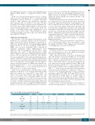
In the cases reported in the literature, the use of thiop- urines was also widespread (63% of all patients with autoimmune diseases and 86% of patients with IBD) (Table 3). TNF-α inhibitors like adalimumab, infliximab and etanercept were, in contrast to our series, the most frequently used drugs in patients with autoimmune dis- eases; 86% was treated a TNF-α inhibitor, often in combi- nation with thiopurines (63% of this group). In the case reports concerning hematologic malignancies most patients were treated with a chemotherapeutic regime containing multiple agents (56%) and a few with chloram- bucil only (6%) (Table 3).
Lymphoma characteristics
An overview of the histological characteristics of the lymphomas of our patients and those reported in the liter- ature is provided in Table 2 and Table 4, respectively. T- NHL were morphologically and immunophenotypically highly variable. In our series, peripheral T-NHL (PTCL- NOS), was seen most frequently (24%), followed by anaplastic lymphoma kinase (ALK)-negative T-cell anaplastic large cell lymphomas (ALCL) (16%). There were three cases (12%) of angioimmunoblastic T-NHL (AITL), limited to the group of patients with autoimmune diseases. We saw only one case of HSTCL. This distribu- tion was comparable to that in the general European pop- ulation.26 However, between the subtype distribution in the reported cases and that in the general population there were some differences. Most notable were the high fre- quency of primary cutaneous T-NHL in the immunocom- promised patients (29% vs. 1.7%), a more frequent occur- rence of HSTCL (12% vs. 1.4%), and the relative lack of AITL cases (1% vs. 18.5%) in this group.26
There was no predominance of either CD4+ or CD8+ lymphomas. Of the 19 cases in which CD4/CD8 staining had been carried out, seven (36.8%) were CD8+ , six (31.6%) were CD4+ , three (15.8%) were CD4+ CD8+ , and three cases (15.8%) were CD4–CD8–. EBER was per- formed in 23 lymphoma cases and the majority was neg- ative (91.3%). EBER was positive in two cases (8.7%), one of which concerned extranodal natural killer (NK)/T-cell lymphoma and the other involving PTCL-NOS with EBV- positive T cells and concurrent EBV-positive B-cell blasts.
In two other cases, both AITL, the malignant T cells were negative, but the B-cell compartment was EBV-positive. Figure 2 shows an example of a PTCL-NOS in a patient with B-cell chronic lymphocytic leukemia (B-CLL) as the underlying disorder.
Extranodal involvement was observed in the vast major- ity of patients (92%), and 16 patients (64%) showed an exclusively extranodal localization of the lymphoma. The most commonly involved organs were the bone marrow or bone, skin, liver, spleen, small bowel and lung. The heart, pancreas, peripheral blood, pelvic cavity organs and central nervous system were affected in some cases (Table 2). This rate of extranodal involvement is higher than the 65-72% which has been reported in the general, immuno- competent population,25,28 and is consistent with more fre- quently occurring extranodal localizations of B-cell lym- phomas in patients with either primary or acquired immunodeficiencies.3,17,29-31
The majority of patients had Ann Arbor stage III/IV dis- ease at presentation (64%). The four patients staged according to the MFCG TNM staging system of cutaneous T-cell lymphomas had stage I or II disease.
Treatment and outcome
As shown in Table 5, in our series lymphoma treatment was very heterogeneous due to the different histological subtypes and clinical stages of disease.
After a median follow-up of six years, 15 patients had died. The median overall survival (OS) was 11.3 months (table 2) and the 1- and 5-year survival rates were 47% (95% confidence interval (CI) 27-67%) and 31% (95% CI 7-55%), respectively. This is somewhat lower than the 5- year survival rate reported in patients with T-NHL in the general population (38-49%),25,28,32 and consistent with a worse survival of HIV and post-transplant patients with T- NHL.14,15,17-19 Since worse outcomes have been reported for certain histological subtypes as compared to others,25,26,28 outcomes were calculated separately for the total number of patients with PTCL-NOS, AITL and HSTCL (n=10). The median OS in this group was 7.5 months with a 5- year survival rate of 20% (95% CI -6-46%), which is indeed lower than the median OS of 60 months and the 5- year survival rate of 41% (95% CI 8-74%) in patients with the remaining histological subtypes.
Twelve of the 15 deceased patients died within four months following diagnosis. The main causes of death, in
Table 3. Use of drugs in cases reported in the literature.
AID
HM
Other ID
Nr pts TNF-αi
IBD 64 60 OtherAID 79 63 Total 143 123 Nrpts CT CLL 44 20
Thiop TNF-αi + thiop MTX CsA Other
55 48 2 - - 35 30 11 6 5 90 78 13 6 5
CA only Melfalan None NR 3 - 13 8 OtherHM 11 9 - 1 1 - Total 55 29 3 1 14 8
None 9
AID: autoimmune disease; CA: chlorambucil; CLL: chronic lymphocytic leukemia; CsA: cyclosporine A; CT: chemotherapy, multiple agents; HM: hematologic malignancies; IBD: inflammatory bowel disease; ID: immunodeficiencies ; MTX: methotrexate; Nr pts: number of patients; Thiop: thiopurines; TNF-αi: TNF-α inhibitors.
haematologica | 2018; 103(3)
491
