
Page 110 - Haematologica3
P. 110
470
X. Wang et al.
cells (Figure 2A, bottom panel). Taken together, our data show that Ara-C induces mouse primary B cells preferen- tially arrested in the S phase of the cell cycle.
Contrary to our findings in primary B cells, in CH12 cells we found that Ara-C treatment caused arrest in the G1 phase at an early time point (6 h: untreated 38.2% ver- sus Ara-C 63.5%) and in the G2/M phase at a later time point (24 h: untreated 10.5% versus Ara-C 42.7%) (Figure 2B, top panel). To further corroborate our findings, we determined the cell cycle progression of G1XP lym- phomas upon Ara-C treatment. Consistently, our data showed that Ara-C also arrested G1XP lymphomas in the G1 phase at an early time point and in the G2/M phase at the later time point (Figure 2B, bottom panel). Ara-C treat- ment resulted in apoptosis regardless of G1 or G2 phase of the cell cycle (Online Supplementary Figure S4A). All cells of the sub-G1 phase were positive for cleaved caspase3, indi- cating their apoptotic phenotypes (Online Supplementary Figure S4B). Nevertheless, the ratio among G1/S/G2 remained the same with or without sub-G1 phase includ- ed. Since we were more interested in living cells, sub-G1 phase cells were not included in the cell cycle analysis.
To distinguish whether Ara-C induces G2 or M phase arrest, we determined the phosphorylation level of histone3 (pH3), which serves as a marker for M phase entry since chromosome condensation requires H3 phosphorylation. We found that pH3 level was reduced upon Ara-C treat- ment, demonstrating that Ara-C-treated lymphomas were arrested in the G2 phase but failed to progress into the M
phase (Online Supplementary Figure S5). These observations are consistent with our findings that the level of pCDK1 (Tyr15) was increased upon Ara-C treatment (Figure 1B), which would inactivate CDK1 and block M phase entry.
In sharp contrast to our findings in CH12 and G1XP, we found that Ramos cells, a human Burkitt lymphoma line, were preferentially arrested in the G1 phase of the cell cycle (Figure 2C). Thus, Ara-C induces differential cell cycle arrest in different types of B cells, namely, S phase arrest in primary mouse B cells, G2 phase arrest in CH12 and G1XP lymphoma cells and G1 arrest in Ramos cells.
p53 deficiency does not affect Ara-C-induced cell cycle arrest or M phase blockage
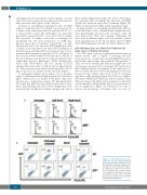
The TP53 gene is the most frequently mutated gene in human cancers.31 p53 is capable of inducing cell cycle arrest, apoptosis, or senescence, modulating DNA repair or metabolism, and serving as the guardian of the genome.32-34 Thus, we tested whether p53 deficiency might affect cell cycle progression upon Ara-C treatment. We treated wt or p53 conditional knock-out primary B cells with Ara-C (1 μM or 10 μM). Our data showed that primary B cells were arrested in the S phase after 24 h of Ara-C treatment regardless of p53 genotype (Figure 3A). Next, we sought to determine whether p53 deficiency affects M phase entry. The percentage of the pH3-positive population is relatively low in primary B cells given that they do not proliferate as fast as lymphomas (Figure 3B, Untreated). In order to increase the percentage of M phase cells, we used col-
B
A
Figure 3. p53 deficiency does not affect the cell cycle changes induced by Ara-C treatment in primary B cells. Wt and p53 conditional knockout pri- mary B cells were treated with 1 μM or 10 μM Ara-C for 24 h. (A) Cell cycles were determined by propidium iodide (PI) staining and (B) mitotic entry was determined by pH3 and PI staining. Data are representative results of three independent experiments.
haematologica | 2018; 103(3)
