
Page 84 - 2020_07-Haematologica-web
P. 84
O.Steinberg-Shemer et al.
consanguinity.1-5 The clinical phenotype includes congeni- tal anomalies, aplastic anemia, a high risk of malignancies, and extreme sensitivity to cross-linking agents. Patients with FA classically present with multiple congenital anomalies and cytopenias, although several patients have no physical defects and normal blood counts, complicat- ing the diagnosis, which is based on the chromosomal breakage test. Genotyping confirms the diagnosis and allows for proper genetic counseling. Some patients exhib- it mosaicism, requiring testing by chromosomal breakage and genetic analysis in non-hematopoietic tissue.6
FA is usually inherited in an autosomal recessive fash- ion, with the exception of the X-linked FANCB and the dominantly inherited FANCR. To date, mutations in more than 20 genes have been detected. Detecting genotype- phenotype correlations is important for prognostic predic- tions, treatment decisions and establishment of directed follow-up programs. However, to date, a clear correlation between the affected gene and the patient's phenotype has not been found. One exception is the more pro- nounced cancer predisposition in patients with FANCD1/BRCA2 and FANCN/PALB2 mutations, in which early onset of malignancy is almost invariably present. Some evidence suggests that the type of mutation corre- lates better with the phenotype than the specific gene. For example, one report found that patients with null muta- tions in FANCA present with a more severe phenotype than those with mutations leading to altered FANCA pro- tein production.7 Conversely, in a separate report, no func- tional or clinical difference was found between patients with absent FANCA protein or those with altered FANCA protein.8 It is possible that specific mutations, as opposed to affected gene or type of mutation, best correlate with the phenotype.9 However, even for a specific mutation, there is a variable phenotypic severity among different ethnicities and among siblings, even twins, suggesting a role for genetic or epigenetic modifiers and/or environ- mental factors.
Here, we present data regarding 111 patients with FA in Israel. This large cohort is unique due to Israel's ethnic diversity, a high degree of consanguinity and a very high percentage of genetically diagnosed patients.
Methods
FA was defined by an abnormal chromosomal breakage test and/or a genetic diagnosis of biallelic mutations in one of the known FA genes. Patients with FA were registered by their treat- ing hematologist as part of the Israeli inherited bone marrow fail- ure registry (I-IBMFR). The I-IBMFR is approved by each local Institutional Review Board. Data was collected at entry to the reg- istry and annually. Data were extracted by the treating physician or by the research team from the patients' charts including demo- graphics, clinical characteristics, laboratory data (including chro- mosomal breakage tests results), molecular diagnosis, and data regarding treatment.
BMF was defined by one of these criteria: a patient who under- went hematopoietic stem cell transplantation (HSCT) for a non- malignant indication, transfusion dependence or at least one cytopenia defined as: absolute neutrophil count (ANC) <1000/μL, platelet count <100,000/μL or hemoglobin <10 g/dL. Severe BMF was further defined as ANC <500/μL and platelet count <20,000/μL.
A five-item congenital abnormality score (CABS) was calculated
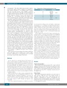
Table 1. Characteristics of Fanconi anemia patients in Israel.
46
62
N= 108 patients
26 10 9
1
54 6 2
Ethnicities
Sephardic
Ashkenazi Jewish
Mixed
Ethiopian
Muslim
Druze Arab
Christian
for each patient by adding up the total number of phenotypic abnormalities in a set consisting of developmental delay, heart or lung abnormalities, renal anomalies, hearing loss and head abnor- malities.10 Whenever possible, genetic analysis was performed as part of the routine work-up. Patients were included in this study if they were clinically suspected as having FA and had either a con- firmed genetic diagnosis of FA or an abnormal chromosomal breakage test, or both.
Genetic analysis was performed by Sanger sequencing, as pre- viously described.11 Sequencing was performed on an ABIPrism 3130xl Genetic Analyzer (Applied Biosystems, Foster City, CA, USA). Chromatograms were visualized with CHROMAS (v.2.6.4; www.thechnelysium.com.au). Variant pathogenicity was deter- mined based on the American College of Medical Genetics and Genomics (ACMG) criteria. Multiplex ligation dependent probe amplification (MLPA) was performed using the commercially available kit SALSA MLPA probemix P031-B2/P032-B2 FANCA (MRC-Holland) following the manufacturer’s instruction.
Data organization was performed with Microsoft Excel (Windows, Version 16.11.1). The data were analyzed using BMDP software (1993, University of California Press, USA). Pearson's chi-square test or Fisher’s exact test (two-tailed) was used for analysis of between-group differences in discrete variables, and analysis of variance (ANOVA) was used for continuous variables. Those variables which did not have Gaussian distributions or when the sample size was very small were compared using the non-parametric Mann-Whitney test. The Kaplan-Meier estimate was used to show survival for the cohort and various sub-groups. Calculations and graphic representation of survival curves were performed on Prism 7 (Graph Pad Software) and on MedCalc soft- ware (Belgium). A P-value of ≤0.05 was considered significant.
Results
Patient demographics
One hundred and eleven patients (53% male) with FA diagnosed between 1980 and 2016 were registered in the I-IBMFR and followed for a median of 15 (range: 0.1-49) years. (Table 1). The median survival time was 27.9 years Brookmayer-Crowley 95% Confidence interval [CI]: 24- 35) (Figure 1A).
Ethnic Origin
Over half of the patients with FA in our cohort were of Arab descent, while the rest were Jewish, mostly of Sephardic origin (Table 1). 63% of the patients were off- spring of consanguineous parents. Consanguinity was reported in 93% of Arab patients and in 21% of Jewish patients. Notably all of the Druze patients were offspring of consanguineous parents.
1826
haematologica | 2020; 105(7)
