
Page 52 - 2020_07-Haematologica-web
P. 52
A. Tiede et al.
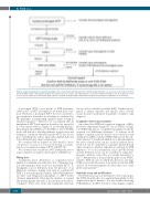
Figure 1. Diagnostic pathway for acquired hemophilia A. The activated partial thromboplastin time (APTT) mixing study will not be needed in an environment in which factor VIII (FVIII) activity is immediately available. Note that the presence of lupus anticoagulant does not exclude acquired hemophilia A. See the ‘Diagnosis’ section for more details. FVIII:C: factor VIII activity; AHA: acquired hemophilia A; ELISA: enzyme-linked immunosorbent assay; rpFVIII: recombinant porcine factor VIII.
A prolonged APTT is not specific to FVIII deficiency; other causes of APTT prolongation are much more com- mon. Therefore, a prolonged APTT does not constitute a good predictive biomarker for bleeding, as evidenced by UK guidelines recommending against routine testing in unselected patients.17 However, we recommend that an unexplained APTT prolongation should not be ignored if it is encountered before surgery or in bleeding patients. Measuring normal FVIII, factor IX (FIX) or factor XI (FXI) levels will exclude a bleeding diathesis in such cases and testing for conditions that prolong the APTT but do not pose a bleeding risk, such as lupus anticoagulant (LA) and factor XII deficiency, may be performed.
We recommend that the diagnosis of AHA should be consid- ered whenever an acute or recent onset of bleeding is accompa- nied by an unexplained prolonged APTT (GRADE 1B).
We recommend that unexplained APTT prolongation prior to surgery should be investigated and not ignored (GRADE 1C).
Mixing tests
Coagulation factor deficiencies or coagulation factor inhibitors, including autoantibodies, LA, or pharmacolog- ical anticoagulants, may result in a prolonged APTT. To distinguish a factor deficiency from the presence of an inhibitory substance, mixing tests may be conducted if FVIII:C is not immediately available. AHA FVIII inhibitors are time- and temperature-dependent, so APTT results obtained immediately following the mixture of normal and patient plasma and after a 2 h incubation should be compared.18 These tests are poorly standardized and can-
not be used to establish or exclude AHA.19 Further investi- gation is always required, and specific factor activity assays should be performed in parallel to facilitate early diagnosis.
Coagulation factor measurements
An isolated low FVIII level suggests a diagnosis of AHA. However, differential diagnoses of low FVIII:C include von Willebrand disease, congenital hemophilia A and the acquired von Willebrand syndrome.19 A decrease in all intrinsic coagulation factors may be an in vitro false result arising from inhibitor-induced FVIII depletion in the sub- strate plasma.20 LA-induced inhibition of phospholipid in the factor activity assay can also result in reduced factor levels. LA can be excluded by a negative diluted Russell viper venom test, which is typically not affected by FVIII inhibitors.21 Vice versa, interference of LA on FVIII activity and the Bethesda assay can be excluded by using chro- mogenic substrate assays that are usually insensitive to LA.22,23 Alternatively, a normal chromogenic assay FVIII:C excludes AHA in cases in which LA decreases one-stage FVIII assay results. However, it should be noted that AHA and LA are both autoimmune disorders that can co-exist in the same patient.24,25
Bethesda assay and modifications
The Bethesda assay was developed to detect and quan- tify FVIII alloantibodies in congenital hemophilia A that display linear type 1 kinetics.26 It is also useful in detecting FVIII inhibitors in AHA, but these often display complex
1794
haematologica | 2020; 105(7)
