
Page 26 - 2020_07-Haematologica-web
P. 26
E. Hellström-Lindberg et al.
with IPSS low/intermediate-1 risk MDS with an age- and sex-matched reference population.19 MDS patients report- ed moderate/severe problems in the dimensions pain/dis- comfort (50%), mobility (41%), anxiety/depression (38%), and usual activities (36%). Limitations were more frequent in older patients, in females, and in those with a high comorbidity burden or needing red blood cell trans- fusions. Finally, Efficace and co-workers studied patients with higher-risk MDS and concluded that patient-report- ed outcomes provide important information regarding the prognosis of patients.20
Disease pathogenesis
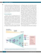
A hallmark of MDS is the dysregulated hematopoietic differentiation resulting in impaired differentiation, mor- phological dysplasia, and cytopenia.63 The cell of origin of MDS lies within the hematopoietic stem and progenitor cell compartment and can usually be tracked back to the pluripotent hematopoietic stem cell, implying that MDS is a malignancy for which cure usually cannot be reached with treatments other than allogeneic stem cell transplan- tation (SCT).21 MDS cells accumulate in the bone marrow as a result of a complex interplay between genetic and epi- genetic alterations, the bone marrow microenvironment, and the immune system, a process that can develop over several years (Figure 1).
The genetic landscape of MDS is quite well delineated. Early studies focused on structural cytogenetic abnormali- ties, identified by metaphase karyotyping in around 50%
of MDS patients. Most of these abnormalities are unbal- anced changes resulting in loss or gain of a large amount of chromosomal material e.g. deletion (del) 5q, monosomy 7, trisomy 8 and del 20q.22 The advent of next-generation sequencing technology resulted in a comprehensive map- ping of the MDS genome.23-25 More than 50 genes have been identified as recurrently mutated in MDS. These genes are involved in biological processes such as DNA methylation, chromatin modification, RNA splicing, cohe- sion formation, regulation of transcription, signaling and DNA repair (Table 4). Some mutations result in specific phenotypes e.g. SF3B1 and del5q which are described below. Interestingly, some of the recurrently mutated genes e.g., DNMT3A, TET2 and ASXL1, are also found in healthy individuals (clonal hematopoiesis of indeterminate progno- sis, CHIP), representing pre-leukemic clones with an age- associated incidence and a varying risk of subsequent development of MDS or other myeloid malignancies.26,27
Several of the recurrently mutated genes are epigenetic regulators.28,29 The MDS epigenome exhibits distinct pathological patterns, which may be explained in part by such mutations but which can also be a consequence of stochastic epigenetic drift, seen with increasing age.30 In analogy with the epigenetic profile, patients with MDS also demonstrate specific gene expression profiles.31-33 Such clusters can be observed for morphological sub- groups e.g. MDS with ringed sideroblasts (MDS-RS) and MDS with excess blasts, as well as for specific genetic lesions e.g., del(5q) and SF3B1.
Many studies have addressed the composition and func- tion of the immune system in MDS and several immuno-
Figure 1. Pathogenesis of myelodyspastic syndromes: underlying mechanisms. CMP: common myeloid progenitors; GMP: granulocyte-monocyte progenitor; MEP: megakaryocyte-erythrocyte progenitor; MkP: megakaryocyte progenitor; EPP: early erythroid progenitor.
1768
haematologica | 2020; 105(7)
