
Page 170 - 2020_07-Haematologica-web
P. 170
P. Strati et al.
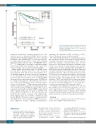
RCHOP & SUVmax ≤18 RCHOP & SUVmax >18
≤18 >18
P=0.02 P=0.001
Others & SUV max
Others & SUV max
Figure 3. Association between overall survival (OS) and maximum standardized uptake (SUVmax) separated by frontline treatment arm.
explaining the difference in OS according to SUVmax observed in patients treated with this regimen.
We acknowledge that there are some limitations due to the retrospective nature of this study and the well-known variability and limited reproducibility of SUV measure- ments.45 For example, consistent criteria may not have been used to select patients in whom to perform a tissue biopsy for exclusion of transformation, and therefore patients with concurrent undiagnosed FL grade IIIB and/or DLBCL may have been included in the study. There was also likely variability in dose-intensity of the chemothera- py between patients. The total metabolic tumor volume (TMTV) was not calculated in this analysis, and the latter has been shown to predict the outcome after frontline therapy in patients with high-burden FL.46 Upon further validation, TMTV and other PET-based functional param- eters, such as total lesion glycolysis, may in the future pro- vide a more standardized approach to assess the prognos- tic value of pre-treatment PET in patients with FL.
Nevertheless, the significant decrease in the CR rate, PFS, and OS observed in patients with SUVmax >18 in our study suggest that a pre-treatment PET scan has a prog- nostic and predictive value in patients with advanced stage FL receiving frontline treatment and prospective ran- domized trials are warranted to investigate this further.
Funding
This research is supported in part by the MD Anderson Cancer Center Support Grant P30 CA016672.
within 24 months from treatment initiation) was over- come by the use of frontline R-CHOP. These novel and clinically relevant findings highlight the need to examine the impact of pre-treatment SUVmax on outcomes in recent- ly completed randomized phase 3 trials in FL comparing different frontline regimens such as R-CHOP, BR, and/or R2.24,25,39 It is important to note that, as only three patients with FL grade IIIA were included in this study, these results can apply only to patients with grades I-II FL.
Finally, despite the beneficial effect of R-CHOP on CR rate and PFS of patients with SUVmax >18, subsequent transformations were still observed among patients treat- ed with this regimen. However, this was not unexpected as not all patients with occult or histologically proven transformed FL achieve durable remissions with R-CHOP. Recurrent disease with transformation after R-CHOP can be salvaged with high-dose chemotherapy with autolo- gous stem cell transplantation and/or chimeric antigen receptor T-cell therapy.40-44 While patients with transfor- mation at the time of relapse who are anthracycline-naïve can be salvaged with R-CHOP, the superior OS observed in patients treated with frontline R-CHOP compared with other regimens within the sub-group of patients with SUVmax >18 (8- year OS 70% vs. 50%, P=0.15) raises the possibility that upfront treatment with anthracycline- based regimen may lead to better outcomes in such patients. On the other hand, relapses occurring in patients with high SUVmax after frontline R-CHOP may have a more aggressive biology, including transformation,
References
1. Jerusalem G, Beguin Y, Najjar F, Hustinx R, Fassotte MF, Rigo P, et al. Positron emission tomography (PET) with 18F-fluo- rodeoxyglucose (18F-FDG) for the staging of low-grade non-Hodgkin's lymphoma (NHL). Ann Oncol. 2001;12(6):825-830.
2. Karam M, Novak L, Cyriac J, Ali A, Nazeer T, Nugent F. Role of fluorine-18 fluoro- deoxyglucose positron emission tomogra- phy scan in the evaluation and follow-up of patients with low-grade lymphomas. Cancer. 2006;107(1):175-183.
3. Wohrer S, Jaeger U, Kletter K, et al. 18F-flu- oro-deoxy-glucose positron emission
tomography (18F-FDG-PET) visualizes fol- licular lymphoma irrespective of grading. Ann Oncol. 2006;17(5):780-784.
4. Elstrom R, Guan L, Baker G, et al. Utility of FDG-PET scanning in lymphoma by WHO classification. Blood. 2003;101(10):3875- 3876.
5. Wirth A, Foo M, Seymour JF, Macmanus MP,
1912
haematologica | 2020; 105(7)
