
Page 127 - 2020_07-Haematologica-web
P. 127
molecules that convey danger signals, which are cumula- tively known as damage-associated molecular patterns (DAMPs).1-4 The spatiotemporally regulated emission of DAMPs by cells undergoing immunogenic cell death (ICD) generates a pronounced immunostimulatory milieu that, in the presence of adequate antigenicity (such as that conferred to cancer cells by somatic mutations), supports the initiation of tumor-targeting immunity.2,5 ICD-relevant DAMPs encompass endoplasmic reticulum (ER) chaper- ones such as calreticulin (CALR, best known as CRT) and
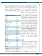
Table 1. Clinical and biological characteristics of acute myeloid leukemia patients.
heat-shock proteins (HSPs), nuclear components such as high mobility group box 1 (HMGB1), nucleic acids, as well as small metabolites like ATP.6,7 In physiological scenarios, DAMPs are mostly intracellular, which prevents their detection by the immune system. Conversely, DAMPs that are secreted into the extracellular space or exposed on the plasma membrane of dying cancer cells can be recog- nized by the immune system via pattern recognition receptors (PRRs), and hence can drive the activation of therapeutically relevant innate and cognate immune responses.2,8 In line with this notion, DAMP accumulation in the tumor microenvironment has been correlated with increased infiltration by multiple immune cell subsets, including mature dendritic cells (DCs) and effector mem- ory T cells.9-12 Moreover, factors linked to danger signaling – including (but not limited to) DAMPs expression levels, PRR expression levels, genetic polymorphisms in DAMP- or PRR-coding genes, and activation of relevant stress responses in cancer cells – have been attributed prognostic values in several cohorts of patients with cancer.13
Considerable work has been dedicated to elucidate the mechanisms whereby DAMPs affect the phenotype and function of myeloid cells that operate as antigen-present- ing cells (APCs).2,8 On the contrary, little attention has been given to the effects of DAMPs on cells of the innate lymphoid system, such as natural killer (NK) cells, despite the fact that NK cells are emerging as potent players in the control of metastases.14 Indeed, surface-exposed HSP fam- ily A member 1A (HSPA1A, best known as HSP70) pro- motes NK-cell-dependent cytotoxicity in vitro15,16 and in vivo,17 while exosome-associated HSP70 can stimulate NK- cell migration and effector functions.18,19 Similarly, extra- cellular HMGB1 can stimulate NK-cell activity upon bind- ing to Toll-like receptor 2 (TLR2) and TLR4.20 Here, we report that CRT exposure on the surface of malignant blasts from acute myeloid leukemia (AML) patients is associated with improved NK-cell secretory and cytotoxic functions. Mechanistic studies revealed that surface- exposed CRT stimulates NK-cell activity indirectly, through the upregulation of IL-15Rα on myeloid CD11c+CD14high cells. Moreover, CRT exposure on AML malignant blasts also correlates with the upregulated expression of genes coding for type I interferon (IFN), which are also involved in the capacity of DCs to enhance NK-cell effector functions.
Methods
Patients
44 patients diagnosed with AML and treated at the Institute of Hematology and Blood Transfusion in Prague between December 2015 and March 2018 plus six AML patients diagnosed and treated at the Department of Hemato-oncology of the Pilsen Hospital between January 2017 and January 2018 were enrolled in this study. Informed consent was obtained according to the Declaration of Helsinki, and the study was approved by the local ethics com- mittee. The main clinical and biological characteristics of the patients are summarized in Table 1. Induction chemotherapy con- sisted mainly (96%) of seven days cytarabine plus idarubicin or daunorubicin for the first three days (standard “7+3” regimen).
Flow cytometry
Peripheral blood mononuclear cell (PBMCs) isolated from AML patients or C57BL/6 (B6) mice, as well as mouse splenocytes,
Danger signaling to NK cells in AML
Variable
Age at diagnosis < 50 years
≥ 50 years Median (years) Range (years)
Sex Male
Female
White blood cell count at diagnosis < 30.000/mm3
≥ 30.000/mm3
Median (109 cells/L)
Range (109 cells/L)
Blasts in peripheral blood Median (%)
Range (%)
De novo AML Secondary AML
FAB classification M0
M1 M2 M4 M5 M6 MDS
Cytogenetic profile Favorable Intermediate Unfavorable Missing data
Molecular characteristics FLT3-ITD
NPM1 mutated
CEBPA mutated
Induction chemotherapy Daunorubicin + Ara-C (3+7) Idarubicin + Ara-C (3+7) FLAG + Idarubicin
Palliative treatment CR
Consolidation Chemotherapy only HSCT
No consolidation
Cohort (n=50)
23 (46%) 27 (54%) 52 21-73
23 (46%)
27 (54%)
42 (84%) 8 (16%) 6.9 0-402.8
25
0-91
41 (82%) 9 (18%)
1 (2%) 10 (20%) 12 (26%) 7 (14%) 10 (20%) 1 (2%) 8 (16%)
6 (12%) 29 (58%) 8 (16%) 7 (14%)
7 (14%) 12 (24%) 2 (4%)
38 (76%) 10 (20%) 1 (2%) 1 (2%) 40 (80%)
14 (28%) 30 (60%) 6 (12%)
AML: acute myeloid leukemia; AM1-ETO: acute myeloid leukemia 1-ETO fusion pro- tein; CEBPA: CCAAT/enhancer-binding protein alpha; CR: complete remission; FLAG: fludarabine + high-dose cytarabine + granulocyte colony-stimulating factor (G-CSF); FLT3-ITD: fms-like tyrosine kinase 3-internal tandem duplication; HSCT: hematopoietic stem cell transplantation; MDS: myelodysplastic syndrome; NPM1: nucleophosmin 1. FAB: French-American-British.
haematologica | 2020; 105(7)
1869
