
Page 111 - 2020_07-Haematologica-web
P. 111
Src kinases and neutrophil extravasation
ABC
DE
FG
HI
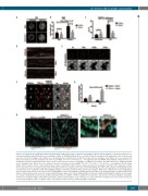
Figure 5. Src family kinases (SFK)-dependent neutrophil elastase (NE) translocation is critical for degradation of the basement membrane. (A) Immunostaining of rep- resentative wildtype and SFK-knockout (ko) neutrophils on BSA- or PECAM-1/ICAM-1/CXCL1–coated (P/I/CXCL1) coverslips for NE analyzed by confocal microscopy. Scale bar: 10 μm. N=3 wildtype and 3 SFK-ko mice. (B) Quantification of ring formation for NE. At least 80 cells from 3 wildtype and 3 SFK-ko mice were analyzed. All data are presented as mean± standard error of the mean (SEM). *P<0.05; ***P<0.001; n.s. : not significant (two-way ANOVA, Sidak multiple comparison test). (C) Quantitative analysis for myeloidperoxidase (MPO) release in blood plasma samples from wildtype and SFK-ko mice 2 hours after TNFα injection by quantitative ELISA assay; 3 wildtype and 3 SFK-ko mice were analyzed. All data are presented as mean±SEM. *P<0.05; **P<0.005; ***P<0.001; n.s. : not significant. (D) NE activity (white) within cremaster muscle venules without (upper image) or with TNFα middle and lower image) stimulation from wildtype and SFK-ko mice imaged by confocal microscopy. Venules were visualized using a CD31 antibody (red) (n=3 mice per group). Scale bar: 40 μm (E) Spinning disk confocal micrographs of LN degradation (indi- cated by black areas compared to white LN staining) by wildtype neutrophils at indicated time points (Lower panels). White lines outline the increase in degradation area over time (Upper panels). Scale bar: 10 μm. (F) Spinning disk confocal micrographs of wildtype (green) and SFK-ko (red) neutrophils plated on antibody-labeled LN (white) at indicated time points. Scale bar: 10 μm. (G) Randomization approach to test for wildtype/SFK-ko interaction strength during migration using the nearest-neigh- bor analysis. (H) Multi photon microscopy of TNFα-stimulated SFK-ko x Lyz2GFP or SFK-ko x Lyz2GFP mice with 1x10⁷ injected deep red labeled wildtype neutrophils. Venules were visualized using a CD31 antibody (turquoise). Dotted squares indicate areas of interest (see panel I) (n=4 mice per group). Scale bar: 30 μm. (I) Close-up of vessel wall of SFK-ko x Lyz2GFP or SFK-ko x Lyz2GFP mice with 1x10⁷ injected deep red labeled wildtype neutrophils.
haematologica | 2020; 105(7)
1853
