
Page 108 - 2020_07-Haematologica-web
P. 108
I. Rohwedder et al.
AB
D
C
E
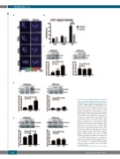
Figure 3. Defective LFA1 clustering and adhesion strengthening in Src family kinases (SFK)-knockout (ko) mice. Integrin downstream signaling was ana- lyzed in isolated wildtype and SFK-knockout (ko) neutrophils. (A) In vitro integrin clustering experi- ments were performed using whole blood from wildtype and SFK-ko mice in flow chambers coated with E-Selectin/ICAM-1 and CXCL1. Blood samples were incubated with anti-LFA1-Alexa547 (2D7) to visualize LFA1 and crawling cells were analyzed for 15 minutes by confocal microscopy. LFA1 intensity is displayed at different time points. White arrows point to the uropod. Color code indicates signal intensity. Dashed lines are exemplary for drawn intensity profiles analyzed in (B) n=3 wildtype and 3 SFK-ko mice. Scale bar: 5 μm. (B) Mean signal intensities of an intensity profile of LFA1 signal, grouped into 3 regions: front, middle and rear of the neutrophil. (C-E) Western blot and respective quan- titative analysis of Syk (Tyr519/520) (72kDa), Paxillin (Tyr118) (68kDa) and Cortactin (Tyr421) (80kDa) phosphorylation after CXCL1 and PMA stimulation in wildtype and SFK-ko neutrophils plat- ed on ICAM1. Band intensity was normalized to respective total protein. All data are presented as mean± standard error of the mean (SEM). *P<0.05; ***P<0.001; n.s. : not significant (one-way ANOVA, Dunnett's multiple comparisons test). N=3 wildtype and 3 SFK-ko mice.
1850
haematologica | 2020; 105(7)
