
Page 10 - 2020_07-Haematologica-web
P. 10
Editorials
Replacing the suppressed hormone: toward a better treatment for iron overload in β-thalassemia major?
Domenico Girelli and Fabiana Busti
Department of Medicine, Section of Internal Medicine, University of Verona, EuroBloodNet Referral Center for Iron Metabolism Disorders, Azienda Ospedaliera Universitaria Integrata of Verona, Verona, Italy
E-mail: DOMENICO GIRELLI - domenico.girelli@univr.it doi:10.3324/haematol.2020.253393
The human body lacks a regulatory mechanism able to excrete excess iron. Therefore, any condition increasing iron entry into the body inevitably results in toxic iron overload.1 The majority of iron over- load disorders can be viewed as endocrine diseases2 caused by insufficient production or activity of hepcidin, the key hormone that finely tunes systemic iron home- ostasis.3 Hepcidin controls body iron content by negative- ly modulating the absorption of dietary iron from the gut,4 and also regulates iron fluxes among different cells and tissues, e.g. from iron-recycling splenic macrophages to erythroid progenitors in the bone marrow.5
Deficiency of this hormone, leading to intestinal iron hyperabsorption, is particularly relevant in the pathogen- esis of hereditary hemochromatosis, due to gene muta- tions impairing hepcidin production, but it is also para- mount in several inherited “iron-loading” anemias,6 par-
ticularly in non-transfusion-dependent thalassemias (NTDT),7 In these conditions, soluble factors produced by erythroid progenitors during expanded/ineffective ery- thropoiesis,8 including erythroferrone,9 directly suppress the synthesis of the hormone in the liver.
Things are more complicated in transfusion-dependent thalassemias (TDT), in which most of the abnormal iron accumulation derives from regular red blood cell transfu- sions, typically every 2-3 weeks.10 Indeed, in TDT, hep- cidin level fluctuates according to suppression of erythro- poiesis by transfusions, with relatively high and low val- ues immediately after and before red blood cell adminis- tration, respectively.11 Thus, increased iron absorption can also contribute to iron overload in TDT, at least during intervals between transfusions.
As for many endocrine disorders, a logical therapeutic approach would be the replacement of the missing hor-
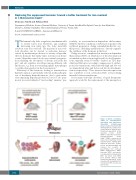
Figure 1. Effects of minihep-
cidins in
dependent and transfusion- dependent β-thalassemia mouse models. Treatment with minihepcidins has similar ben- eficial effects in both the non- transfusion-dependent β-tha- lassemia mouse model (Hbbth3/+) and in the novel trans- fusion-dependent model (Hbbth1/th2BMC) developed by Casu et al. In particular, in the transfused mice, minihepcidins improved ineffective erythro- poiesis and splenomegaly, and
non-transfusion-
reduced the
parenchymal iron overload. ERFE: erythroferrone; HEPC: hepcidin; NTDT: non-transfu- sion-dependent β-thalassemia; RBC: red blood cells; BMC: bone marrow chimera; TDT: transfusion-dependent β-tha- lassemia; Hb: hemoglobin; Ret: reticulocytes; ROS: reactive oxy- gen species.
severity of
1752
haematologica | 2020; 105(5)
