
Page 15 - Haematologica May 2020
P. 15
Editorials
AB
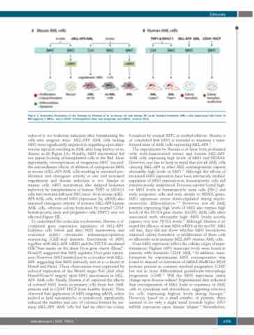
Figure 1. Schematic illustration of the findings by Sharma et al. in mouse (A) and human (B) acute myeloid leukemia (AML) cells expressing high levels of Meningioma 1 (MN1) and in CD34+ hematopoietic stem and progenitor cell (HSPC) controls (Ctrl).
reduced in vivo leukemia induction after transplanting the cells into syngenic mice. MLL-AF9+ AML cells lacking MN1 were significantly impaired in engrafting upon intra- venous injection resulting in AML after long latency or no disease at all (Figure 1A). Notably, MN1 inactivation did not impair homing of transplanted cells to the BM. Most importantly, overexpression of exogenous MN1 'rescued' the anti-leukemic effects of ablation of endogenous MN1 in mouse rMLL-AF9 AML cells resulting in increased pro- liferation and clonogenic activity in vitro and increased engraftment and disease induction in vivo. Similar to murine cells, MN1 inactivation also delayed leukemia induction by transplantation of human THP1 or MV4;11 cells into immune deficient NSG mice. As in mouse rMLL- AF9 AML cells, reduced MN1 expression (by siRNA) also impaired clonogenic activity of primary MLL-AF9 human AML cells, whereas colony formation by normal CD34+ hematopoietic stem and progenitor cells (HSPC) was not affected (Figure 1B).
To understand the molecular mechanisms, Sharma et al. compared gene expression signatures of MLL-AF9+ leukemic cells before and after MN1 inactivation, and evaluated public chromatin immunoprecipitation sequencing (ChIP-seq) datasets. Enrichment of MN1 together with MLL-AF9, MEIS1 and the DOT1L-mediated H3K79me marks on the distal Hoxa gene cluster (Hoxa7- Hoxa10) suggested that MN1 regulates Hoxa gene expres- sion. However, MN1 seemed not to co-localize with MLL- AF9, suggesting that MN1 primarily acts as a co-factor of Hoxa9 and Meis1. These observations were supported by reduced expression of the Hoxa9 target Bcl2 (and other Hoxa9/Hoxa10 targets) upon MN1 inactivation in MLL- AF9+ AML cells. Finally, Sharma et al. explored the effects of reduced MN1 levels in primary cells from five AML patients and in CD34+ HSCP from healthy donors. They observed that application of MN1-targeting siRNA, either packed in lipid nanoparticles or transfected, significantly reduced the number and size of colonies formed by pri- mary MLL-AF9+ AML cells but had no effect on colony
formation by normal HSPC in methylcellulose. Sharma et al. concluded that MN1 is essential to maintain a trans- formed state of AML cells expressing MLL-AF9.11
The experiments by Sharma et al. have been performed with well-characterized mouse and human MLL-AF9+ AML cells expressing high levels of MN1 and HOXA9. However, one has to keep in mind that not all AML cells carrying MLL-AF9 or other MLL rearrangements express aberrantly high levels of MN1.10 Although the effects of increased MN1 expression have been intensively studied, regulation of MN1 expression in hematopoietic cells still remains poorly understood. Previous reports found high- est MN1 levels in hematopoietic stem cells (HSC) and early progenitor cells and very similar to HOXA genes, MN1 expression seems down-regulated during myelo- monocytic differentiation.5,12 However, not all AML patients expressing high levels of MN1 also express high levels of the HOXA gene cluster. Inv(16)+ AML cells often associated with aberrantly high MN1 levels mostly express very low HOXA levels.13 Although Sharma et al. tested the efficacy of anti-MN1-siRNA in the inv(16)+ ME1 cell line, they did not show whether MN1 knockdown impaired colony formation or proliferation of these cells as efficiently as in primary MLL-AF9+ murine AML cells.
Does MN1 expression reflect the cellular origin of trans- formation? Highest MN1 transcript levels were found in patients with immature CD34+ AML.14 In addition, trans- formation by experimental MN1 overexpression was found to depend on activation of MEIS1/AbdB-like HOX proteins present in common myeloid progenitors (CMP) but not in more differentiated granulocyte-macrophage progenitors (GMP).15 Will the MN1 expression status change upon disease relapse? Experimental data indicated that overexpression of MN1 leads to resistance of AML cells to cytarabine and doxorubicin, suggesting selection for cells expressing highest levels during therapy.16 However, based on a small number of patients, there seemed to be only a slight trend towards higher MN1 mRNA expression upon disease relapse.17 Nevertheless,
haematologica | 2020; 105(5)
1175
