
Page 139 - Haematologica May 2020
P. 139
MLL-rearranged AML requires MN1
through Hoxa7 were not significantly dysregulated, while Hoxa9, Hoxa10 and Meis1 were strongly down-regulated in MLL-AF9/Mn1null cells (Figure 6A). We also performed global gene expression analysis in MLL-AF9/Mn1wt ver- sus MLL-AF9/Mn1null cells with significant differentially expressed genes (Online Supplementary Table S6 and Online Supplementary Figure S15A).
To evaluate a direct transcriptional effect of MLL-AF9 and MN1 on the distal Hoxa cluster genes, we analyzed the binding of MN1, MLL-AF9, Hoxa9, MEIS1 and dimethyl marks of H3K79 (representing DOT1L binding) at the Hoxa cluster locus from available chromatin immunoprecipitation-sequencing (ChIP-Seq) data sets.
MN1 chromatin marks were enriched at distal Hoxa clus- ter genes (Hoxa7 to Hoxa10) similar to MLL-AF9, MEIS1 and H3K79me2 marks (Figure 6B), suggesting that MN1 is a direct regulator of Hoxa cluster gene expression. However, genome-wide co-localization of MLL-AF9 and MN1 chromatin did not show any overlap of a significant number of chromatin marks, in contrast to the high over- lap of MN1 with Hoxa9 and MEIS1 (Figure 6C-E), suggest- ing that MN1 is essential for MLL-AF9 primarily due to its role as a co-factor of Hoxa9 and Meis1.
To substantiate this observation, we evaluated the expression of Bcl2 in MLL-AF9/Mn1 wild-type and null cells, as Bcl2 is a known target of Hoxa9 but not of MLL-
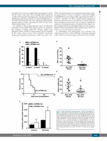
A
B
CD
E
Figure 3. MLL-AF9-induced leukemogenesis requires Meningioma 1 (MN1). (A) Engraftment of MLL-AF9/Mn1wt and MLL-AF9/Mn1null cells in peripheral blood (PB) at the indicated time points [mean±standard error of mean (SEM) of the indicated number of mice]. (B) White blood cell count (WBC) in peripheral blood of mice at four weeks. Mice received transplants of MLL-AF9/Mn1wt and MLL-AF9/Mn1null cells (mean±SEM, n=10). (C) Survival of mice receiving transplants of MLL-AF9/Mn1wt and MLL- AF9/Mn1null cells (log rank test). (D) Spleen weight of the indicated mice at the time of death (mean±SEM, n=10). (E) Homing assay. Percentage of MLL-AF9/Mn1wt and MLL-AF9/Mn1null cells in bone marrow at 8 and 24 hours after transplantation (mean±SEM of the number of mice, n=5 for each cell type and each time point). *P<0.05; **P<0.01; ns: not signifi- cant.
haematologica | 2020; 105(5)
1299
