
Page 48 - Haematologica April 2020
P. 48
J. Perez Botero et al.
chronic, patients can have low hemoglobin, microcytosis and increased red cell distribution width from secondary iron deficiency. Other abnormalities of the complete blood count (CBC), suggest an alternative diagnosis.
Coagulation screening tests
Routine tests ordered in the workup of a patient with abnormal bleeding, such as prothrombin time (PT), acti- vated thromboplastin time (aPTT) and fibrinogen, are usually normal, unless a patient is being evaluated in the setting of significant acute hemorrhage and has evidence of consumptive coagulopathy.
Platelet function screening tests
The platelet function analyzer (PFA)-100 provides a measure of platelet function under high shear. It is con- venient as it uses low volume whole blood samples and is widely available to clinicians. Very prolonged closure times (>300 seconds) are compatible with GT but not specific, as other disorders, such as severe von Willebrand disease, Bernard Soulier syndrome and afibrinogenemia, can produce the same result. However, a normal PFA-100 has a very high negative predictive value for GT and vir- tually excludes this diagnosis.19
Platelet light transmission aggregometry
Despite its limited availability and the need for larger- volume samples and immediate processing, platelet light transmission aggregometry (LTA) remains the gold stan- dard in the clinical diagnosis of GT. It is based on the decreased turbidity generated by platelet agglutinates or aggregates in platelet rich plasma after exposure to differ- ent agonists. Decreased/absent aggregation (<10%) with all physiologic agonists, together with a normal aggluti- nation response to ristocetin (mediated by GPIb-IX-V), is the classic pattern observed in patients with GT.20 Due to the large variability in platelet aggregation results and the significant effect of pre-analytical variables on this test, confirmation of the findings in a second sample is recom- mended.
Whole blood impedance aggregometry
This is available in several centers worldwide. While it can be performed in whole blood samples and using lower volumes, there is not enough evidence to support equivalent sensitivity and reproducibility when com- pared to LTA.21 There is some clinical utility to this test in cases in which access to LTA is difficult; however, patients should preferably be referred for evaluation at a center that has LTA capacity at least once to confirm the diagnosis.
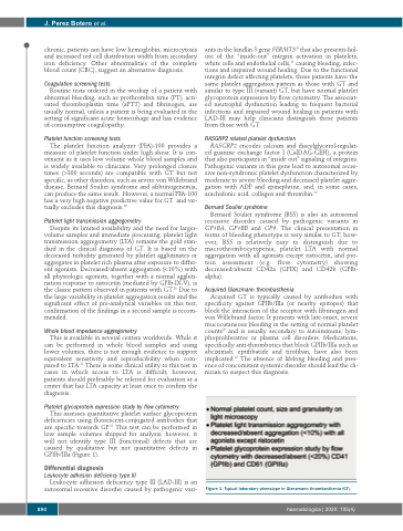
Platelet glycoprotein expression study by flow cytometry
This assesses quantitative platelet surface glycoprotein deficiencies using fluorescent-conjugated antibodies that are specific towards GP.22 This test can be performed in low sample volumes shipped for analysis; however, it will not identify type III (functional) defects that are caused by qualitative but not quantitative defects in GPIIb/IIIa (Figure 1).
Differential diagnosis
Leukocyte adhesion deficiency type III
Leukocyte adhesion deficiency type III (LAD-III) is an autosomal recessive disorder caused by pathogenic vari-
ants in the kindlin 3 gene FERMT323 that also presents fail- ure of the “inside-out” integrin activation in platelets, white cells and endothelial cells,11 causing bleeding, infec- tions and impaired wound healing. Due to the functional integrin defect affecting platelets, these patients have the same platelet aggregation pattern as those with GT and similar to type III (variant) GT, but have normal platelet glycoprotein expression by flow cytometry. The associat- ed neutrophil dysfunction leading to frequent bacterial infections and impaired wound healing in patients with LAD-III may help clinicians distinguish these patients from those with GT.
RASGRP2 related platelet dysfunction
RASGRP2 encodes calcium and diacylglycerol-regulat- ed guanine exchange factor I (CalDAG-GEFI), a protein that also participates in “inside out” signaling of integrins. Pathogenic variants in this gene lead to autosomal reces- sive non-syndromic platelet dysfunction characterized by moderate to severe bleeding and decreased platelet aggre- gation with ADP and epinephrine, and, in some cases, arachidonic acid, collagen and thrombin.24
Bernard Soulier syndrome
Bernard Soulier syndrome (BSS) is also an autosomal recessive disorder caused by pathogenic variants in GP1BA, GP1BB and GP9. The clinical presentation in terms of bleeding phenotype is very similar to GT; how- ever, BSS is relatively easy to distinguish due to macrothrombocytopenia, platelet LTA with normal aggregation with all agonists except ristocetin, and pro- tein assessment (e.g. flow cytometry) showing decreased/absent CD42a (GPIX) and CD42b (GPIb- alpha).
Acquired Glanzmann thrombasthenia
Acquired GT is typically caused by antibodies with specificity against GPIIb/IIIa (or nearby epitopes) that block the interaction of the receptor with fibrinogen and von Willebrand factor. It presents with late-onset, severe mucocutaneous bleeding in the setting of normal platelet counts25 and is usually secondary to autoimmune, lym- phoproliferative or plasma cell disorders. Medications, specifically anti-thrombotics that block GPIIb/IIIa such as abciximab, eptifibatide and tirofiban, have also been implicated.26 The absence of lifelong bleeding and pres- ence of concomitant systemic disorder should lead the cli- nician to suspect this diagnosis.
Figure 1. Typical laboratory phenotype in Glanzmann thrombasthenia (GT).
890
haematologica | 2020; 105(4)
