
Page 310 - Haematologica April 2020
P. 310
M. Hetzel et al.
improved total protein, cholesterol, GM-CSF, M-CSF and MCP-1 levels in the BALF twelve weeks after secondary HSC-GT (Figure 5A-G). These data, in combination with the low transgene levels and VCN observed in the PB and BM of the primary recipients (Figure 3F), suggested that only some of the secondary recipients engrafted with long-term reconstituting HSC. This was supported by VCN analysis in secondary recipients, which revealed that those mice not benefitting from the transplantation had
A
no or barely detectable VCN in their BM (VCN 0 and 0.03), in contrast to the mice presenting with an improved PAP phenotype (VCN 0.46 and 1.93) (Figure 5H). These data clearly indicate that the disease phenotype can be ameliorated in secondary recipients, in which engraftment of gene-corrected cells can be achieved.
In addition to secondary transplantation, we also per- formed long-term studies in primary recipients. Here, 5 of 7 mice presented AM in the range of 8 to 64% of all
B
C
DEF
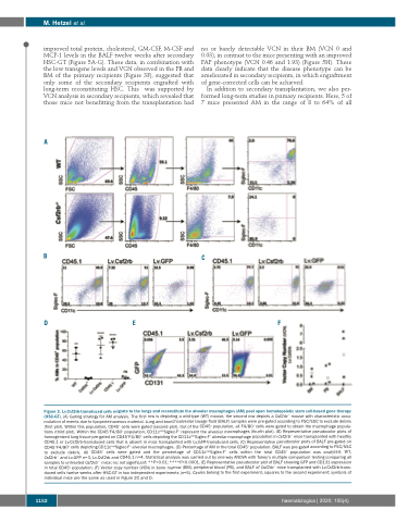
Figure 3. Lv.Csf2rb-transduced cells migrate to the lungs and reconstitute the alveolar macrophages (AM) pool upon hematopoietic stem cell-based gene therapy (HSC-GT). (A) Gating strategy for AM analysis. The first row is depicting a wild-type (WT) mouse, the second row depicts a Csf2rb-/- mouse with characteristic accu- mulation of events due to lipoproteinaceous material. Lung and bronchoalveolar lavage fluid (BALF) samples were pre-gated according to FSC/SSC to exclude debris (first plot). Within this population, CD45+ cells were gated (second plot). Out of the CD45+ population, all F4/80+ cells were gated to obtain the macrophage popula- tions (third plot). Within the CD45+F4/80+ population, CD11chighSiglec-F+ represent the alveolar macrophages (fourth plot). (B) Representative pseudocolor plots of homogenized lung tissue pre-gated on CD45+F4/80+ cells depicting the CD11chighSiglec-F+ alveolar macrophage population in Csf2rb-/- mice transplanted with healthy CD45.1 or Lv.Csf2rb-transduced cells that is absent in mice transplanted with Lv.GFP-transduced cells. (C) Representative pseudocolor plots of BALF pre-gated on CD45+F4/80+ cells depicting CD11chighSiglec-F+ alveolar macrophages. (D) Percentage of AM in the total CD45+ population. BALF was pre-gated according to FSC/SSC to exclude debris. All CD45+ cells were gated and the percentage of CD11chighSiglec-F+ cells within the total CD45+ population was analyzed. WT, Csf2rb-/- and Lv.GFP n=3, Lv.Csf2rb and CD45.1 n=4. Statistical analysis was carried out by one-way ANOVA with Tukey’s multiple comparison testing comparing all samples to untreated Csf2rb-/- mice; ns: not significant; **P<0.01; ****P<0.0001. (E) Representative pseudocolor plot of BALF showing GFP and CD131 expression in total CD45+ population. (F) Vector copy number (VCN) in bone marrow (BM), peripheral blood (PB), and BALF of Csf2rb-/- mice transplanted with Lv.Csf2rb-trans- duced cells twelve weeks after HSC-GT in two independent experiments (n=5). Circles belong to the first experiment, squares to the second experiment; symbols of individual mice are the same as used in Figure 2C and D.
1152
haematologica | 2020; 105(4)
