
Page 18 - Haematologica April 2020
P. 18
Editorials
ed (i.e. by surgical intervention) than patients with actual pathological fractures. Substantial differences were observed with less average blood loss, shorter hospital stays, greater likelihood of discharge, and greater likeli- hood of resuming support-free ambulation.6,18,19
If pathological fractures do occur, they are mainly treat- ed surgically to stabilize the fractured bones and to improve patients’ quality of life via pain relief and restora- tion of function and mobility.20 Palliation, not cure, is usu- ally the surgical objective of treatment of MM-related bone disease.
The findings of Thorsteinsdottir et al. deserve the inter- est of interdisciplinary teams of MM experts. However, further investigations are needed to corroborate and better understand these findings, i.e. that the risk of death after suffering a fracture has not significantly decreased com- pared to patients who do not develop a fracture after the introduction of more effective treatment agents in MM. Moreover, subsequent analyses should study the risk of death after fracture in MM patients compared to patients without cancer. This is of particular importance since eld- erly patients in general have a higher morbidity and mor- tality after fractures.
As a consequence of Thorsteinsdottir et al.’s and previ- ous findings, the early recognition of an impending frac- ture remains highly relevant in MM and other cancer patients. This includes taking a detailed general medical history, especially if aggravating or novel pain occurs or reoccurs. The following questions should be asked: “Is there any new onset of pain?” “Is there pain at night?” “Is
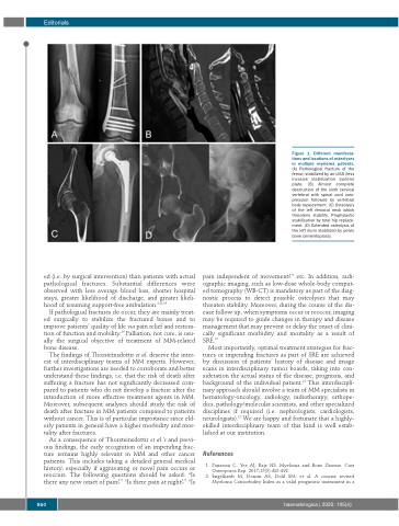
Figure 1. Different manifesta- tions and locations of osteolyses in multiple myeloma patients. (A) Pathological fracture of the femur, stabilized by an LISS (less invasive stabilization system) plate. (B) Almost complete destruction of the sixth cervical vertebral with spinal cord com- pression followed by vertebral body replacement. (C) Osteolysis of the left femoral neck which threatens stability. Prophylactic stabilization by total hip replace- ment. (D) Extended osteolysis of the left ilium stabilized by pelvic bone cementoplasty.
pain independent of movement?” etc. In addition, radi- ographic imaging, such as low-dose whole-body comput- ed tomography (WB-CT) is mandatory as part of the diag- nostic process to detect possible osteolyses that may threaten stability. Moreover, during the course of the dis- ease follow up, when symptoms occur or reoccur, imaging may be required to guide changes in therapy and disease management that may prevent or delay the onset of clini- cally significant morbidity and mortality as a result of SRE.15
Most importantly, optimal treatment strategies for frac- tures or impending fractures as part of SRE are achieved by discussion of patients’ history of disease and image scans in interdisciplinary tumor boards, taking into con- sideration the actual status of the disease, prognosis, and background of the individual patient.21 This interdiscipli- nary approach should involve a team of MM specialists in hematology-oncology, radiology, radiotherapy, orthope- dics, pathology/molecular scientists, and other specialized disciplines if required (i.e. nephrologists, cardiologists, neurologists).22 We are happy and fortunate that a highly- skilled interdisciplinary team of this kind is well estab- lished at our institution.
References
1. Panaroni C, Yee AJ, Raje NS. Myeloma and Bone Disease. Curr Osteoporos Rep. 2017;15(5):483-498.
2. Engelhardt M, Domm AS, Dold SM, et al. A concise revised Myeloma Comorbidity Index as a valid prognostic instrument in a
860
haematologica | 2020; 105(4)
