
Page 135 - Haematologica April 2020
P. 135
Clofazimine inhibits chronic myeloid leukemia
Clofazimine reduces MYB expression by rapid degradation of p65 NFκB protein
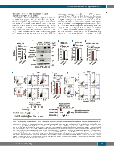
Clofazimine reduced MYB mRNA expression from 1 h in K562 cells (Figure 4B); we therefore investigated whether it regulates any factor that regulates MYB expres- sion itself. A literature search revealed that NFκB tran- scription factors regulate MYB expression by various mechanisms.34-37 Consistent with a reported NFκB response element (NFκB-RE) in the MYB promoter situat- ed at -278 to -256 bp upstream of the transcriptional start site,37 tumor necrosis factor-α treatment, or p65/RELA
AB
transfection activated a MYB (-687-+204) promoter reporter38 (Figure 5A). Given that clofazimine downregu- lated PRDX1 promoter luc in HEK-293 cells (Figure 2L) and that p65 is endogenously expressed in HEK-293 cells,39 we investigated whether clofazimine regulates p65 expres- sion. Clofazimine reduced p65 protein in K562 cells with- in 15 min, without affecting other NFκB family members, p50, p105 and C-Rel (Figure 5B). Clofazimine did, howev- er, fail to alter p65 mRNA expression (Figure 5C) indicat- ing that clofazimine-mediated p65 downregulation may happen at a post-transcriptional or -translational level.
CDE
FG
H
IJ
Figure 3. Introduction of exogenous PRDX1 in cells compromises clofazimine-induced reactive oxygen species generation, apoptosis and differentiation. (A-D) Overexpression of peroxiredoxin 1 (PRDX1) in K562 cells ameliorates clofazimine (CFZ)-induced reactive oxygen species generation (A), caspase cleavage and BAX expression (B), apoptosis (C; representative dot plots in Online Supplementary Figure S6A), and CD61 expression (D; representative histograms in Online Supplementary Figure S6B). (E-H). CD34+ chronic myeloid leukemia (CML) cells transfected with PRDX1 are protected from CFZ-induced ROS generation and apop- tosis. (E, F) CD34+ cells were isolated from chronic phase (CP)-CML cells as described above and were transfected with empty vector or a PRDX1 expression plasmid. Cells were then treated with CFZ (5 μM; 24 h) and dihydroethidium fluorescence was measured by flow cytometry (E; graphical representation, F; representative dot plots). (G, H) CD34+ cells transfected with empty vector or PRDX1 were treated with CFZ (5 μM; 48 h) and apoptosis was assessed by annexin V staining followed by flow cytometry (G; graphical representation, H; representative dot plots). (I) PRDX1 mRNA expression in CP-CML cells determined by quantitative real-time poly- merase chain reaction (QRT-PCR). (J) PRDX1 mRNA expression in CD34-38+, CD34+38+ and CD34+38- cells by QRT-PCR. Graphs (except I & J) are mean ± standard error of mean of three independent experiments. Immunoblots are representative of three independent experiments. **P<0.01, ***P<0.001 (A,C,D-E,G; two-way analysis of variance followed by the Bonferroni post-test. I,J; Kruskal-Wallis test followed by the Dunn test). DCFDA: 2',7'-dichlorofluorescein diacetate; V: vehicle; SSC; side scatter; DHE: dihydroethidium; PE: phycoerythrin.
haematologica | 2020; 105(4)
977
