
Page 98 - Haematologica March 2020
P. 98
A. Caulier et al.
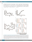
preferentially expressed in CD34+ cells and in early stages of erythropoiesis from day 4 to 10 (corresponding to burst-forming unit-erythroid/colony-forming unit-ery- throid/proerythroblast in our culture system) then decreased during terminal maturation (Figure 1A). This was in agreement with previously published RNA- sequencing analyses on erythroid precursors.14,23,24
AB
D
C
Figure 1. PIEZO1 expression during human in vitro erythroid differentiation. PIEZO 1 expression was assessed at day 4 in CD45low/CD123-/CD34+/CD36- cells, and at day 7 in CD36+ cells, for both the gene and protein expression experiments. (A) PIEZO1 mRNA expression (determined by quantitative reverse transcriptase poly- merase chain reaction, RT-qPCR) relative to HPRT expression, during synchronized erythroid differentiation. Differential expression relative to day 0. Statistical analy- sis was made compared to day 10. No significant change was seen at days 4, 7, and 12. (B) Glycophorin A (GPA) mRNA expression (determined by RT-qPCR) relative to HPRT expression, during synchronized erythroid differentiation. Reference was day 0. (C) Kinetics of relative PIEZO1 protein expression during in-vitro erythroid differentiation, in parallel to relative GPA membrane expression. For both, expression at each time point was assessed by multiparametric flow cytometry (MFC) (mean fluorescence intensity at the time point relative to that at day 10.) (D) MFC histograms of PIEZO1 protein expression assessed at different culture time points (red). We used both the secondary antibody alone (blue) and a non-specific rabbit anti HLA-DR1 antibody (orange) as controls. (n=3 for all experiments). ***P<0.001; **P<0.01; * P<0.05.
Expression of glycophorin A (GPA) was measured in par- allel as a positive marker of erythroid differentiation (Figure 1B). Similar results were observed at the protein level using MFC (Figure 1C, D). The specificity of PIEZO1 antibody staining using MFC was verified by western blot and immunofluorescence assays (Online Supplementary Figure S4).
612
haematologica | 2020; 105(3)
