
Page 89 - Haematologica March 2020
P. 89
ELANE knockout restores granulopoiesis in congenital neutropenia
ELANE knockout in HSPC of ELANE-CN patients restores diminished granulocytic differentiation
To further evaluate the clinical applicability of ELANE KO as a treatment option of ELANE-associated CN, we performed CRISPR/Cas9 RNP-mediated gene editing in primary bone marrow CD34+ HSPC of four ELANE-CN patients (Table 1) and three healthy donors and differenti- ated the cells towards neutrophils. ELANE knockout in CD34+ HSPC was performed by electroporation of human CD34+ HSPC with assembled ELANE specific sgRNA and Cas9 protein (Figure 5A). The editing efficiency varied between 27 % and 94 % (Figure 5B, Online Supplementary Figure S7). As expected, NE levels in neutrophils differen- tiated from the total population of edited cells were markedly reduced (Figure 5C-D, Online Supplementary Figure S8A-B). Moreover, ELANE KO leads to elevated granulocytic differentiation, as assessed by the percentage of CD15+CD11b+CD45+ cells (Figure 6A, Online Supplementary Figure S9A-B) and morphological examina- tion of cytospin preparations of mature granulocytes gen- erated on day 14 of the in vitro granulocytic differentiation using liquid culture (Figure 6B). At the same time, the ratio of ELANE KO cells increased from day 7 to day 14 of dif- ferentiation (Figure 5B). Simultaneously, the percentage of CD34+CD45+ cells was reduced in ELANE KO cells of CN patients, but not in healthy donor cells (Online Supplementary Figure S10A). In one patient (CN I120F), no difference in the percentage of CD15+CD11b+CD45+ cells between MOCK and ELANE KO samples was observed, but a clear improvement of granulocytic differentiation was detected in cytospin slides. This finding may be explained by relative mild neutropenia (Online Supplementary Table 3) and possible expression of CD15 in not fully mature myeloid cells in this patient.
Scanning and transmission electron microscopy revealed that ELANE KO cells of both healthy control and one CN patient showed no significant differences in mor- phology or intracellular structures, compared with MOCK cells of a healthy donor (Figure 6C-D).
Altogether, these data suggest that ELANE KO cells have a differentiation advantage over the HSPC carrying mutat- ed ELANE.
MOCK and ELANE KO neutrophils generated from a healthy donor or one CN patient (Figure 7C).
Chemotactic activity of fMLP-treated neutrophils was also comparable between MOCK and ELANE KO groups (Figure 7D).
A
Neutrophils generated from ELANE KO HSPC exhibited unaffected ROS production, phagocytosis and chemotaxis upon activation in vitro
We further evaluated in vitro activation of neutrophils gen- erated from ELANE KO HSPCs in liquid culture for 14 days. We first performed an assessment of H2O2 levels (ROS) in fMLP-activated ELANE KO neutrophils generated from a healthy donor. We detected no differences between ELANE WT and ELANE KO neutrophils (Figure 7A).
Phagocytosis was evaluated by incubation of cells with fluorescein-conjugated Staphylococcus aureus BioParticles for two hours. Percentage of GFP+ granulocytes that engulfed bacteria were assessed by FACS using gating on granulocyte population in the dot plot of forward-scatter light (FSC) versus side-scatter light (SSC) channels. We did not detect any significant differences in phagocytosis of ELANE KO neutrophilic granulocytes, as compared to control MOCK cells (Figure 7B). As an independent evalu- ation of phagocytosis kinetics, we performed live cell imaging of neutrophils incubated with pHrodo Green E. coli Bioparticles Conjugate using IncuCyte ZOOM sys- tem and observed similar phagocytosis behavior of
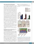
Figure 4. ELANE knockout restored granulocytic differentiation of ELANE-CN iPSC. (A) Flow cytometry analysis of suspension cells harvested from embryoid body (EB)-based granulocytic cell culture of respective iPSC clones on day 28 or 32 of differentiation. Data represent means ± standard deviation (SD) from two independent experiments. *P<0.05, **P<0.01. (B) Wright-Giemsa staining of cytospin preparations of suspension myeloid cells harvested from iPSC culture at day 28 or 32 of differentiation. Representative images are depicted. (C)
B
C
+
Colony-forming unit (CFU) assay of CD34 cells harvested from EB-based iPSC
culture on day 14 of differentiation. Data represent means ± SD from two inde- pendent experiments. *P<0.05.
haematologica | 2020; 105(3)
603
