
Page 86 - Haematologica March 2020
P. 86
M. Nasri et al.
CRISPR/Cas9-gRNA RNP mediated ELANE KO in iPSC and HSPC
Electroporation was carried out using the Amaxa nucle- ofection system (P3 primary kit, #V4XP-3024) according to the manufacturer’s instructions. 1x10⁶ human iPSC or CD34+ HSPC were electroporated with assembled gRNA (8 mg) and Cas9 (15 mg) protein (Integrated DNA Technologies).
Isolation of single cell iPSC clones
8 x 10³ human iPSC were plated on Geltrex-coated 10- cm dish in StemFlex medium (Thermo Fisher Scientific, #A3349401) and RevitaCell supplement (Thermo Fisher Scientific, #A2644501). The medium was changed every 24 hours without RevitaCell supplement. On day 7, single iPSC colonies were picked and transferred to the Geltrex- coated 96-well plates (one clone/well).
Colony Forming Unit (CFU) assay
CD34+ cells were resuspended in IMDM supplemented with 2 % FBS (Stemcell Technologies, #07700) and enriched Methocult (Stemcell Technologies, #H4435). The cell suspension was plated on 3.5 cm dishes (3x10³ cells/dish) for 14 days.
A
In vitro phagocytosis assay
Cells were incubated with or without fluorescein-conju-
gated Staphylococcus aureus BioParticles (Invitrogen, #S2851) at a ratio of 100 particles per cell for two hours at 37°C, washed twice with PBS/ 2 % BSA, resuspended in 300 mL FACS buffer and analyzed by flow cytometry.
Statistical analysis
Differences in mean values between groups were ana- lyzed using two-sided, unpaired Student’s t-tests using GraphPad Prism software.
Additional Material and Methods are available in the Online Supplementary Material and Methods.
Results
Inhibition of ELANE expression restored defective granulocytic differentiation of HL60 cell lines express- ing endogenous ELANE mutations
We created CRISPR/Cas9 edited mutant ELANE knock- in HL60 human promyelocytic cell lines expressing either p.P139L or p.C151Y ELANE mutations. All-trans retinoic acid (ATRA) induced differentiation of wild-type and
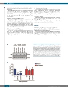
Figure 1. The effect of ELANE knock-down on the impaired myeloid differentiation of HL60 cells expressing mutant neutrophil elastase. (A) CRISPR/Cas9 edited human promyelocytic HL60 cells expressing p.P139L and p.C151Y mutant neu- trophil elastase (NE) were electroporated with scrambled and anti-ELANE siRNA and maintained in the complete medium for five days. Western blot (WB) analysis at day 4 shows complete knock-down of NE detected with an anti-NE monoclonal antibody. For loading control, the membrane was stripped and re-probed with a β-actin-specif- ic antibody. Representative WB membranes are depicted. (B) Myeloid differentiation was induced with 2 mM ATRA (all-trans retinoic acid). After five days, cells were labeled with CD11b myeloid differentiation surface marker and examined using Fluorescence-activated cell sorting (FACS) analysis. The proportion of CD11b-PE pos- itive cells is indicated. Data represent means ± SD from four independent experi- ments. Two-sided, unpaired Student’s t-test P-values are shown, ***P<0.0001 and ***P=0.0043 for p.P139L and p.C151Y respectively compared to wild-type (WT).
B
600
haematologica | 2020; 105(3)
