
Page 50 - Haematologica March 2020
P. 50
P. Pignatelli et al.
rectional. Therefore, patients carrying aPL have an increased risk of ischemic heart disease, and conversely, after a first coronary artery disease event, patients positive for aPL showed twice the risk of recurrent major adverse cardiovascular events at 12 and 24 months.13 This risk was also evident in subjects with juvenile myocardial infarc- tion in absence of cardiovascular risk factors.13
Concerning obstetric complications, fetal loss, especially after 10th week of gestation and premature birth due to eclampsia or placental insufficiency are frequent complica- tions in APS women.14 In young women with a history of multiple miscarriage, the immunological study for aPL should be considered.
Finally, catastrophic APS (C-APS) is a severe and life- threatening manifestation characterized by simultaneous venous or/and arterial thrombosis, often triggered by infections and surgical procedures. C-APS involves multi- ple organs and systems due to excess of proinflammatory cytokines, coagulation cascade, and platelet activation, leading to thrombosis and microangiopathic hemolytic anemia.15
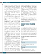
In addition to the above described signs and symptoms, the clinical presentation of patients with APS may be more heterogeneous, involving thrombosis of medium and small vessels (Table 1).6,7,16-18 The relevance in clinical practice of non-conventional APS criteria was investigated during the 14th Congress on Antiphospholipid Antibodies, in which each relevant clinical manifestation was ana- lyzed and each evidence was evaluated by the GRADE system. This system also considers the balance of patient- important outcomes, the overall quality of the evidence for each outcome, and any uncertainty about values.18
The most commonly affected sites are the kidney, the skin, and the cardiovascular and nervous systems. In the kidney, it is possible to find an acute thrombotic microangiopathy or a chronic pattern of vaso-occlusive lesions such as cortical ischemic lesions, arterial fibrous intimal hyperplasia or interstitial fibrosis.6 APS nephropathy can be identified with a urine test associat- ed with a 24-hour investigation of proteinuria. A biopsy is mandatory in cases where the cause is not clearly iden- tifiable, as in patients with concomitant diabetes, uncon- trolled arterial hypertension or other autoimmune dis- eases such as SLE.
Concerning the skin, livedo reticularis can be found, and recurrent ulcerations called livedoid vasculopathy have also been described.8 To evaluate skin abnormalities, a clinical examination is often adequate, and there is usually no need for skin biopsy.
Cardiac abnormalities include valve leaflet thickening,6 and diastolic dysfunction, especially of the right ventri- cle.19 Heart valve disease and diastolic dysfunction can be investigated by resting transthoracic echocardiography and by cardiac magnetic resonance imaging (MRI) if a myocardial involvement is suspected (i.e. myocarditis). Pericardium may also be involved, especially in patients with APS and SLE.
Finally, APS is associated with an increased risk of dementia, seizures, multiple sclerosis-like illness, migraine, myelitis transversa and chorea, due to vascular damage and a direct action of antibodies on neurons and ependymal cells.7,20,21 However, unlike other neurological disorders, seizure is not considered a non-conventional criterion due to lack of strength of evidence.18 To identify critical illness such as neurological disorders, instrumental
examinations are mandatory. An MRI and an electroen- cephalogram could be useful in recognizing brain atrophy associated with dementia and seizures, and could identify more elusive symptoms such as chorea and migraine if associated with an accurate physical examination.
Other blood alterations include thrombocytopenia (commonly mild with platelet count between 50x109/L and 150x109/L, but also severe with platelet counts <20x109/L often associated with microangiopathy) and hemolytic anemia with the possible presence of schisto- cytes.6 In particular, thrombocytopenia is common in APS, affecting 20-46% of patients and could paradoxically be associated with an increased risk of thrombosis.18 Thrombocytopenia may be the result of an increased acti- vation and destruction of platelets by an immune-mediat- ed mechanism involving aPL or by thrombotic microan- giopathy.22
After the exclusion of a pseudo-thrombocytopenia, and performing a Coombs test to ascertain the autoimmune nature of thrombocytopenia, corticosteroids, immunosup- pressive agents, immunoglobulins and new drugs such as Mammalian target of rapamycin (mTOR) inhibitors and monoclonal antibodies could be helpful in patients with autoimmune thrombocytopenia.23
Definition of seronegative antiphospholipid syndrome and non-criteria antiphospholipid antibodies
The first definition of SN-APS was given in 2003 by Hughes and Khamashta5 who described patients with clin- ical manifestations highly suggestive of APS in absence of the laboratory criteria such as LAC, aCL and aβ2GPI anti- bodies.
Seronegative APS is usually a diagnosis of exclusion and should be suspected in patients with a clinical history sug- gestive of APS, such as those with recurrent arterial venous thrombotic events, recurrent miscarriage, or unexplained thrombocytopenia, with persistent negativity of aPL tested on at least two occasions, and when other causes of throm-
Table 1. “Extra-criteria” manifestations of antiphospholipid syndrome.
Nervous system
Dementia
Seizures
Multiple sclerosis–like illness Chorea
Myelitis
Skin
Livedo reticularis Livedoid vasculopathy
Heart
Valve vegetations or thickening (Libman-Sacks Endocarditis) Diastolic dysfunction
Blood Thrombocytopenia Hemolytic anemia
Kidney
Microangiopathy
Chronic vaso-occlusive lesions (atherosclerosis, glomerular
ischemia, interstitial fibrosis, arterial fibrous intimal hyperplasia)
564
haematologica | 2020; 105(3)
