
Page 276 - Haematologica March 2020
P. 276
H. de Boussac et al.
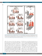
Figure 5. Kinase inhibitors enhance the sensitivity of multiple myeloma cells to conventional treatments. Human myeloma cell lines (HMCL) were cultured for four days in 96-well flat-bottom microtiter plates in RPMI 1640 medium, 10% fetal calf serum, 2 ng/mL IL-6 culture medium (control) and graded Melphalan concentra- tions (A) or Lenalidomide concentrations (B) in presence or absence of IC20 of CHK1i, MELKi, PBKi, CDC7-DBF4i, SRPKi, MPS1i and PLK4i. IC50 were calculated after viability assessment by CellTiter-Glo® Luminescent Cell Viability Assay. Results are representative of three independent experiments. P-value: *<0.05; **<0.01; ***<0.001. S: significant synergy calculated by the method of Chou and Talalay.
MPS1i did not increase cell death (Online Supplementary Figure S9C and S10A). Next, we monitored DNA damage by measuring levels of the DNA double-strand break (DSB) marker γH2AX after the different co-treatments. As expected, Melphalan treatment alone, even at the sub- lethal dose, increased the level of γH2AX, while Lenalidomide did not demonstrate any effect (Figure 6B and Online Supplementary Figure S10B). However, among all the combinations tested, only MELKi significantly potentialized Melphalan-induced DNA damage in AMO1 but not in OPM2 cells. Interestingly MELKi, CDC7-DBF4i and SRPK1i alone induced DSB as monitored by γH2AX levels (Figure 6B and Online Supplementary Figure S9D) although it should be noted that high concentrations of the CHK1 inhibitor AZD7762 or MELK inhibitor
OTSSP167 induced early DSB that progressively decrease as monitored by measuring γH2AX in AMO1 after 24 and 48 hours of treatment (Online Supplementary Figure S11). Thus, the significant potentialization of Melphalan and Lenalidomide toxicity by CHK1i, MELKi, CDC7-DBF4i and SRPK1i appears to be due to an increased induction of apoptosis, and not to an increase of DNA damage or cell cycle deregulation (Online Supplementary Figure S12).
According to these results, we investigated the thera- peutic interest of kinases inhibitors to overcome Melphalan resistance using Melphalan resistant (Mres) XG7 and XG2 cell lines (Figure 7A and Online Supplementary Figure S13A). Interestingly, while no clear differences could be observed for the IC50 of MELKi, CHK1i, PBKi and MPS1i in the Mres and sensitive (WT)
AB
790
haematologica | 2020; 105(3)
