
Page 191 - Haematologica March 2020
P. 191
Targeting BCL2 and MAPK in AML
proliferation and promote differentiation, rather than induce death.33,37,38 Remarkably, over 60% of patients’ sam- ples responded to the combination therapy, notably including samples that were insensitive to both agents on their own. Moreover, these responders carried diverse
genetic alterations that affect leukemia cell proliferation (FLT3, RAS), differentiation (RUNX1), genomic stability (NPM1), and epigenetic modifications (TET2, IDH1 and IDH2). Clonogenic assays demonstrated that the combi- nation markedly impaired the colony-forming functions
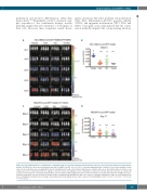
AB
CD
Figure 4. In vivo administration of cobimetinib in combination with venetoclax demonstrated anti-leukemia efficacy in acute myeloid leukemia xenograft mouse models. (A) NSGS mice were injected intravenously with OCI-AML3-Luci-GFP cells (1.0×10⁶). Leukemia engraftment was confirmed 1 week later through a nonin- vasive in vivo bioluminescence imaging (BLI) system following injection with a D-luciferin (4 mg/mouse) substrate. Mice were dosed daily with oral vehicle or an orally active form of cobimetinib (Cobi; 10 mg/kg) or venetoclax (Ven; 100 mg/kg) or their combination (Combo) for 4 weeks. BLI data over time are shown. (B) Luciferase intensity [mean ± standard deviation(SD)] at week 5. Human CD45 engraftment in bone marrow and spleen was determined by time-of-flight mass spec- trometry (C) BLI data over time from the leukemia model established with MOLM13-Luc-GFP cells (1×10⁶ per animal) in NSGS mice. Mice received treatment as for the OCI-AML3/Luc/GFP model for 14 days. (D) Quantification of BLI signals (mean ± SD) on day 17 in the MOLM13 model. *P<0.05; **P<0.01; ***P<0.001; ****P<0.0001.
haematologica | 2020; 105(3)
705
