
Page 154 - Haematologica March 2020
P. 154
T. Sun et al.
A
B
D
E
C
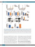
Figure 6. Insufficient action of the WDR4–IL-6 axis decreases hematopoiesis-supportive activities of bone marrow derived mesenchymal stromal cells (BM-MSC) from patients with JAK2V617F-positive ET. A–B. Numbers of BFU-E, CFU-E, CFU-GM, CFU-Mix, and CFU-MK formed by purified normal CD34-positive cells after cocul- ture with BM-MSC infected with LV-shWDR4, or LV-WDR4. WDR4 increased the number of BFU-E, CFU-GM, and CFU-Total formed by normal CD34-positive cells (A). No changes were observed in the number of CFU-MK (B). C. WDR4 increased the secretion of IL-6 from BM-MSC as determined by ELISA on the supernatant obtained from the MSC cultures. D–E. WDR4 increased the intracellular expression of IL-6 in BM-MSC as determined by Western blotting (D) and qPCR (E). MSC used in each assay were at passage four. All the experiments were repeated at least three times. *P 0.05; **P<0.01; ***P<0.001; ****P<0.0001. Data are presented as the mean ± SD. qPCR: quantitative real-time polymerase chain reaction; CFU-E: colony-forming unit-erythroid; CFU-GM: colony-forming unit-granulocyte and macrophage; CFU-Total: total colony-forming units; CFU-MK: colony-forming unit-megakaryocyte; n: number of unique donors in each group; ns: not significant; NC: normal control; SD: standard deviation.
MSC infected with LV-shWDR4, the numbers of erythroid burst-forming units (BFU-E), CFU-GM, and CFU-Total were significantly lower than those in the control groups (Figure 6A). In contrast, increasing WDR4 expression cor- rected the ET MSC-mediated defects, as evidenced by higher numbers of BFU-E, CFU-GM, and CFU-Total rela- tive to those in the control groups (Figure 6A). No changes were observed in the CFU-MK number (Figure 6B).
We next examined whether WDR4 regulated the hematopoietic cytokines mentioned above that were differ- entially produced between the two MSC samples. The results revealed a link between IL-6 and WDR4 (Figure 6C). We next performed qPCR and Western blotting to assess
whether IL-6 expression was affected by WDR4. In WDR4 knock-down HD MSC, intracellular IL-6 protein (Figure 6D) and mRNA levels (Figure 6E) were decreased. Furthermore, in the ET MSC, restoration of WDR4 expression alleviated the decrease in IL-6 levels (Figure 6 D–E). Taken together, these results indicate that WDR4 promotes the intracellular expression and secretion of IL-6 by BM-MSC.
WDR4 acts through the ERK–GSK3β–CREB pathway to increase IL-6 expression and secretion by BM-MSC
To identify candidate kinases through which WDR4 may act on IL-6, we evaluated the levels of 43 phosphory- lated kinases in HD MSC infected with LV-shWDR4 rela-
668
haematologica | 2020; 105(3)
