
Page 41 - 2020_02-Haematologica-web
P. 41
Innate immune cells in sickle cell disease
investigations are required to confirm the role of dendritic cells and the BMP/SMAD signaling pathway in SCD bone complications.
Neutrophils
Neutrophils have long been suspected to be involved in the pathophysiology of SCD. The absolute neutrophil count is higher in SCD patients in steady-state than in eth- nicity-matched healthy controls and is positively correlat- ed with SCD severity.27 A high leukocyte count is also a risk factor for early death, acute chest syndrome (ACS), hemorrhagic stroke and sickle nephropathy.28-31 Conversely, decreased neutrophil count may have positive effects, as suggested by a report of an alleviated SCD phe- notype in a patient with associated congenital neutropenia who experienced the first episodes of VOC after the intro- duction of granulocyte colony-stimulating factor (G-CSF) to treat neutropenia.32 Thus, G-CSF and granulocyte- macrophage colony-stimulating factor (GM-CSF) should be strictly avoided in SCD patients because myeloid growth factors are responsible for VOC and ACS.33,34 Hydroxyurea may have clinical benefit for SCD patients even in the absence of elevated fetal hemoglobin (HbF) level, but a decrease in neutrophil count is always observed in such cases, and hydroxyurea seems most effective in patients with the greatest reduction in neu- trophils.35
In addition to these quantitative aspects, several studies have highlighted the activated state of neutrophils from
SCD patients, with increased adhesive properties at base- line and even more during VOC.36,37 Clinical manifesta- tions of SCD have been found to be associated with the expression of adhesion molecules on leukocytes.38 Hydroxyurea may also benefit SCD patients by suppress- ing neutrophil activation and correcting the dysregulated expression of adhesion markers on these cells.39,40 An important step in understanding the role of neutrophils in SCD pathophysiology was the intravital microscopy demonstration in SCD mice of increased neutrophil adhe- sion to the endothelium but also to sickle RBC in postcap- illary venules.41 In this model, mice deficient in endothelial P-selectin and E-selectin displayed defective leukocyte recruitment to the vessel wall and were protected against vaso-occlusion. E-selectin was found to induce a second- ary wave of activating signals, resulting in the clustering of activated macrophage-1 antigen (Mac-1) on the leading edge of adherent neutrophils, thereby allowing for the capture of sickle RBC and platelets. Here again, inactiva- tion of E-selectin or Mac-1 prevented neutrophil–RBC and neutrophil–platelet interactions, thereby improving blood flow in the microcirculation and mouse survival.42 These findings suggest that interactions between activated endothelium, activated neutrophils, captured sickle RBC and platelets can contribute to decreased blood flow, fur- ther accentuating RBC sickling, neutrophil recruitment and tissue ischemia.43 In blood samples from SCD patients, studied in microfluidic flow chambers, neu- trophils rolling on E-selectin under shear stress were found
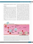
Figure 1. Monocytes in sickle cell disease. Interaction of sickle red blood cells (RBC) with endothelial cells enhances cellular oxidant stress, resulting in increased transendothelial migration of blood monocytes. Suspected mechanisms of monocyte activation in sickle cell disease (SCD) involve hypoxemia, platelet-monocyte aggregates mediated by P-selectin/P-selectin glycoprotein ligand 1 interaction, RBC/reticulocyte-monocyte interactions, placental growth factor released from RBC and co-stimulation of toll-like receptor 4 by heme and lipopolysaccharide. SCD activated monocytes display increased expression of CD1, CD11b and tissue factor on their surfaces, as well as increased production of interleukin-1β and tumor necrosis factor-α. In turn, activated monocytes activate endothelial cells through the nuclear factor-κB pathway, resulting in enhanced expression of intercellular adhesion molecule 1, vascular cell adhesion molecule 1 and E-selectin. Patrolling mono- cytes uptake cellular debris derived from heme-exposed endothelial cells, thus leading to high expression of heme oxygenase-1. Patrolling monocytes also scavenge endothelium-adherent sickle RBC. HO-1: heme oxygenase-1; ICAM-1: intercellular adhesion molecule 1: IL1-β: interleukin 1 beta; LPS: lipopolysaccharide; Lu/BCAM: Lutheran/basal cell adhesion molecule; NF-κB: nuclear factor kappa B; PlGF: placental growth factor; PSGL-1: P-selectin glycoprotein ligand 1; TF: tissue factor; TLR4: toll-like receptor 4; TNF-α: tumor necrosis factor alpha; VCAM-1: vascular cell adhesion molecule 1.
haematologica | 2020; 105(2)
275
