
Page 230 - 2020_02-Haematologica-web
P. 230
A. Natoni et al.
ical role of sialylation in melanoma metastasis and growth in vivo.32
The vehicle-pre-treated MM1SHeca452 cells showed an ini- tial response to bortezomib in vivo. Indeed, bortezomib was able to reduce tumor burden, however, despite this initial response, bortezomib alone was not able to improve survival. These data would suggest that the sur- viving MM1SHeca452 cells were so aggressive that they still induced death in mice at a similar rate to the non-borte- zomib-treated mice. 3Fax-Neu5Ac pre-treatment of MM1SHeca452 cells effectively blocked α2-3 and α2-6 sialyla- tion as well as expression of SLea/x. More importantly, pre- treatment of MM1SHeca452 cells with 3Fax-Neu5Ac blunted
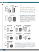
Figure 5. 3Fax-Neu5Ac treatment reduces motility of MM1SHeca452 independently of SDF1α. MM1SHeca452 cells were treated with 300 μM 3Fax-Neu5Ac or dimethyl sulfoxide (DMSO) (vehicle control) for seven days. After treatment, cells were starved for 1 hour (h) and then seeded on the upper chamber of transwells. Lower chamber was filled with either serum-free media (No SDF1α) or serum- free media supplemented with SDF1α (20 ng/mL). Cells were allowed to migrate for 4 h at 37°C. After incubation, cells in the lower chambers were col- lected and counted using a BD Accuri flow cytometer. Data are presented as (A) raw data or (B) as the difference between migrating cells in SDF1α-containing media and control media. Bars represents mean±Standard Error of Mean of three independent experiments. One-way ANOVA test was used to determine statistical significance with Sidak’s multiple comparison post-hoc testing. *P<0.05; **P<0.01; ***P<0.001; ns: non-significant.
ABC
DEF
G
Figure 6. 3Fax-Neu5Ac treatment impairs adhesion and rolling of MM1SHeca452 on E-selectin, VCAM1 and MADCAM1 under shear stress. MM1SHeca452 cells were treated with 300 μM 3Fax-Neu5Ac or dimethyl sulfox- ide (DMSO) (vehicle control) for seven days. After treatment, cells were collected, washed and resuspended at 2x10⁶/mL. Eighty μL of cell suspension were loaded onto E-selectin- (A-C), MADCAM1- (D-F) and VCAM1- (G) coated microfluidic channels and adhesion/rolling assay was performed at 0.5 dyne/cm2 at RT using the Mirus Evo NanoPump. Rolling cells were imaged using an A-Plan 10X/0.25 objective of an A10 Vert.A1 microscope equipped with a QIClick F-M-12 Mono camera. Images were acquired using the Vena Flux Assay software and analyzed using the Image-Pro Premiere. Bars represent the mean±Standard Error of Mean of three independent experiments. Unpaired Student’s t-test was used to determine statistical sig- nificance. *P<0.05; **P<0.01; ns: non-significant.
464
haematologica | 2020; 105(2)
