
Page 23 - 2020_02-Haematologica-web
P. 23
Editorials
Insights into the composition of stroke thrombi: heterogeneity and distinct clot areas impact treatment
Rui-Gang Xu and Robert A. S. Ariëns
Discovery and Translational Science Department, Leeds Institute of Cardiovascular and Metabolic Medicine, University of Leeds, Leeds,UK
E-mail: ROBERT A. S. ARIËNS - r.a.s.ariens@leeds.ac.uk doi:10.3324/haematol.2019.238816
The structure of blood clots has recently become a hot topic in the scientific and medical literature. A search on PubMed for ‘clot structure’ results in >1,000 publications in the last 25 years, with >400 in the last five years alone. The reason for this surge in studies of the structure of blood clots is that dense clot structures that are resistant to fibrinolysis have been associated with both arterial and venous thrombosis (reviewed by Undas and Ariëns1), and lead to poor outcome in prospective studies of arterial2 and venous3 thrombotic disease. Furthermore, mechanisms from cellular contributions from platelets, red blood cells (RBC)4 and white blood cells (neutrophils producing extracellular traps or NET5) to clot structure are increasingly being understood. However, many previous studies have focused on indi- vidual clot components and their roles in clot structure and function, largely using either in vitro or in vivo method- ology. Holistic approaches to study clot structure in thrombi obtained from patients with thrombosis have until now been few and far between, and are increasingly needed to place studies of individual clot structures into clinical context.
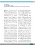
In the current edition of Haematologica, Staessens et al.6 studied the internal organization of 177 thrombi collected from endovascularly treated ischemic stroke patients. The objective of this study was to gain further under- standing of the composition of stroke thrombi by histo- logically analyzing the internal organization of their structural components, including fibrin, RBC, von Willebrand factor (vWF), platelets, leukocytes and DNA. Using bright field and fluorescence microscopy, the authors observed that stroke thrombi are very heteroge- neous in nature in two ways. First, they differ distinctly from each other in size, shape, and color. Second, within each thrombus there is considerable heterogeneity, with different areas or segments of the thrombus demonstrat- ing different structural components. Based on the major structural components that are dominant in the thrombi, they were classified into two distinct types: RBC- and platelet-rich areas of the thrombus (Figure 1). Platelet-rich areas are composed of dense fibrin structures, platelets, vWF, leukocytes and extracellular DNA, whereas compo- sition of RBC-rich areas is less complex, with packed RBC and a thin fibrin meshwork filling the gaps in between the packed RBC as the main structural components. Staessens et al.6 further quantified the relative contribution of each type of thrombus area. It appears that the contri- bution of both types varies significantly across all throm- bi analyzed. For most thrombi, both regions are dispersed throughout, although some thrombi have more clearly defined boundaries, with RBC-rich regions surrounded by platelet-rich regions. The findings obtained in this
study provide interesting insight into the composition and internal organization of stroke thrombi and could be helpful in furthering our understanding of thrombolysis resistance and developing new therapies for acute ischemic stroke. Furthermore, knowledge of the structur- al composition of thrombi may be important for the rela- tive success of mechanical thrombectomy.
This study makes important and valuable contributions to the previous efforts in the identification of thrombi composition, many of which were based on scanning electron microscopy.7-12 There are a number of significant strengths of the current study. First, compared to previous studies,10,11 this is the first study to image thrombi obtained from patients with stroke with such detail, including visualization of multiple structural components of thrombi: fibrin, vWF, DNA and blood cells at the same time. Second, the use of immunofluorescence in this study overcomes limitations of conventional staining and generated stunning images, which allow for accurate localization of specific components and provide impor- tant structural insight of stroke thrombi both at cellular and molecular levels. In particular, compared with previ- ous studies using conventional staining,7,12-14 more detailed organization and structural features of platelet, vWF and fibrin meshes in thrombi were demonstrated. Interestingly, the polyhedral morphology of RBC inside thrombi is also defined in these images, reminiscent of the tightly packed polyhedrocytes previously observed in thrombi formed in vivo and in vitro.15,16 Furthermore, the observation that extracellular DNA and leukocytes were located primarily in the platelet-rich areas, and close to the interface of these areas with others, emphasizes their potential crucial role in the rt-PA resistance observed in patients.
While this study makes big strides forward in our understanding of thrombus structure and function, some limitations remain. The first is that only microscopic methods were used to study the histological composition of thrombi, while there are no data on mechanical or functional properties of the thrombi. Previous studies on mechanical properties have shown that fibrin-rich clots have a higher friction than RBC-rich clots,17 and the increased percentage of RBC in the clot affects fibrin net- work heterogeneity and clot stiffness,18 indicating that differences in thrombi composition may be strongly linked to the mechanical properties of these clots. The mechanical properties of thrombi may in turn impact on the degree of embolization on one hand, and the success rate of thrombus retrieval by endovascular thrombecto- my on the other. A comprehensive study of the mechan- ical properties of thrombi in patients is needed to further our understanding of the possible correlations between
haematologica | 2020; 105(2)
257
