
Page 83 - Haematologica Atlas of Hematologic Cytology
P. 83
CHAPTER 11 - Myelodysplastic/myeloproliferative neoplasms
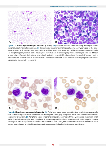
AB Figure 1 Chronic myelomonocytic leukemia (CMML) (A) Peripheral blood smear showing monocytosis with morphologically normal monocytes (B) Bone marrow smear showing high cellularity and hyperplasia of the gran- ulocytic series with predominant intermediate and and late forms and and less than 5% blasts (CMML-0) Erythroid cells are are morphologically normal Some neutrophils have nuclear chromatin pro ections Monocytic cells are are difficult to to appreciate If dysplasia is is is is is absent or minimal as as as in in this case CMML diagnosis can be made if monocytosis is is is is is persistent and all other causes of monocytosis have been excluded or or an an acquired clonal cytogenetic or or molec- ular genetic abnormality is present AB Figure 2 Chronic myelomonocytic leukemia (A) Peripheral blood smear revealing two atypical monocytic monocytic cells with with rather clumped nuclear chromatin and finely granulated gray cytoplasm Note also a a a a a a a a a a neutrophil with with hy- pogranular cytoplasm (B) Peripheral blood smear showing promonocytes with finely dispersed chromatin small nucleoli and abundant light blue cytoplasm A promonocyte differs from a a a a a a a a monoblast for the irregular nuclear outline it is is a a a a a a a a a blast blast equivalent and and should be be counted as as as such Thus the distinction between a a a a a a a a a monoblast and and a a a a a a a a a promonocyte has no no practical importance as as as they are regarded as as as having the the same significance 70
