
Page 286 - Haematologica Atlas of Hematologic Cytology
P. 286
ABC
DEF
GHI
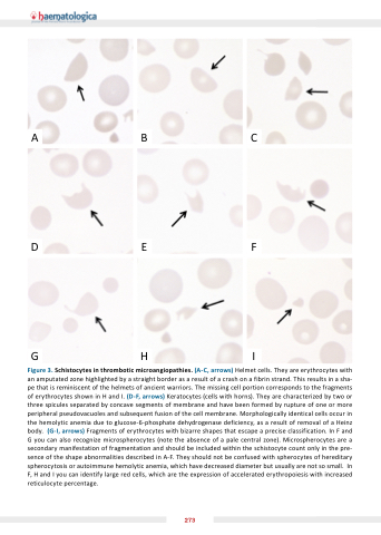
Figure 3 Schistocytes in thrombotic microangiopathies
( arrows) Helmet cells They are erythrocytes with an an amputated zone highlighted by a a a a a a a a a a a a a straight border as as a a a a a a a a a a a a a result result of a a a a a a a a a a a a a crash on on a a a a a a a a a a a a a fibrin strand This results in in a a a a a a a a a a a a a sha- pe that is is is reminiscent of of the the helmets of of ancient warriors The missing cell portion corresponds to the the fragments of erythrocytes shown in H and I
(D F
arrows) Keratocytes (cells with horns) They are characterized by two or or three spicules separated by by concave segments of of membrane and have been formed by by rupture of of one or or or more peripheral pseudovacuoles and subsequent fusion of the cell cell membrane Morphologically identical cells occur in the hemolytic anemia due to glucose-6-phosphate dehydrogenase deficiency as as a a a a a a a a result of of removal of of a a a a a a a a Heinz body ( I
I
arrows) Fragments of erythrocytes with bizarre shapes that escape a a a a a a a a a a precise classification In F
F
and G you can also recognize microspherocytes (note the absence of a a a a a a a a pale central zone) Microspherocytes are a a a a a a a a secondary manifestation of fragmentation and should be included within the the schistocyte count only in in in the the pre- sence of of the shape abnormalities described in A-F They should not be be confused with spherocytes of of hereditary spherocytosis or autoimmune hemolytic anemia which have decreased diameter but usually are not so small In F
H and I
you can identify large red cells which are the expression of accelerated erythropoiesis with increased reticulocyte percentage 273
