
Page 246 - Haematologica Atlas of Hematologic Cytology
P. 246
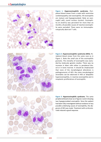
Figure 1 Hypereosinophilic syndrome
Peri- pheral blood smear from a a a a male with restrictive cardiomyopathy and eosinophilia All eosinophils are mature and hypogranulated Note an an an eosi- nophil with round nucleus (center) Eosinophi- lia (>1 5x10⁹/L) was persistent for more than six months all possible causes of reactive eosinophi- lia were were excluded There were were no no immunophe- notypically aberrant T cells Figure 2 Hypereosinophilic syndrome
(HES) Pe- ripheral blood smear from the same case as as in Figure 1 Note the the small size of the the eosinophilic granules The clonality of eosinophils was exclu- ded by molecular genetic studies There was no increase in in in blast cells either in in in peripheral blo- od or in bone marrow It should be emphasized that none of the morphological alterations is pathognomonic of HES: the same morphological anomalies can be observed in HES or idiopathic hypereosinophilia in in in in reactive eosinophilia eosinophilia and in in in in neoplastic proliferations of eosinophils Figure 3 Hypereosinophilic syndrome
The same peripheral blood smear as in in Figures 1 and 2 showing two hypogranulated eosinophils Since the pa ent had been fully inves gated without evidence of any possible underlying causes of eosinophilia the dia- gnosis of HES was made Treatment with cor coste- roids induced a good response 233
