
Page 196 - Haematologica Atlas of Hematologic Cytology
P. 196
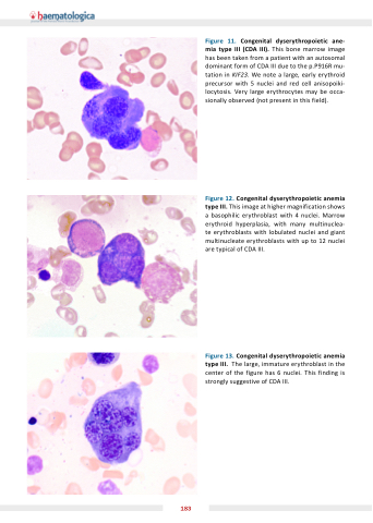
Figure 11. Congenital dyserythropoietic ane- mia type III ( D  III). This bone marrow image has been taken from a patient with an autosomal dominant form of CDA III due to the p.P916R mu- tation in KIF23. We note a large, early erythroid precursor with 5 nuclei and red cell anisopoiki- locytosis. Very large erythrocytes may be occa- sionally observed (not present in this field).
Figure 12. Congenital dyserythropoietic anemia type III. This image at higher magnification shows a basophilic erythroblast with 4 nuclei. Marrow erythroid hyperplasia, with many multinuclea- te erythroblasts with lobulated nuclei and giant multinucleate erythroblasts with up to 12 nuclei are typical of CDA III.
Figure 13. Congenital dyserythropoietic anemia type III. The large, immature erythroblast in the center of the figure has 6 nuclei. This finding is strongly suggestive of CDA III.
183
