
Page 188 - Haematologica Atlas of Hematologic Cytology
P. 188
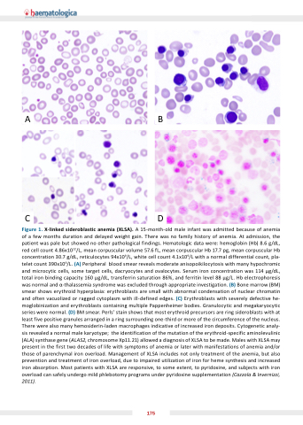
AB
CD
Figure 1 1 lin ed ed sideroblastic anemia anemia ( LS ) A 15-month-old male infant was admitted because of anemia anemia of of a a a a a a a a a a few months duration and delayed weight gain There was no family history of of anemia At admission the patient was pale but showed no other pathological findings Hematologic data were: hemoglobin (Hb) 8 6 g/dL red cell count 4 86x1012/L mean mean mean corpuscular corpuscular corpuscular volume 57 6 6 fL mean mean mean corpuscular corpuscular corpuscular Hb Hb 17 7 7 7 pg mean mean mean corpuscular corpuscular corpuscular Hb Hb concentration 30 7 g/dL reticulocytes 94x109/L white cell count count 4 4 1x10⁹/L with a a a a a normal differential count count pla- telet count 390x109/L ( ) Peripheral blood smear reveals moderate anisopoikilocytosis with many hypochromic and and microcytic cells cells some target cells cells dacryocytes and and ovalocytes Serum iron concentration was 114 g/dL total iron binding capacity 160 g/dL transferrin saturation 86% and ferritin level 88 g/L Hb electrophoresis was was normal and -thalassemia syndrome was was excluded through appropriate investigation ( ( ) ) Bone marrow (BM) smear shows erythroid hyperplasia: erythroblasts are small with abnormal condensation of nuclear chromatin and often vacuolized or ragged cytoplasm with with ill-defined edges ( ) Erythroblasts with with severely defective he- moglobinization and and erythroblasts containing multiple Pappenheimer bodies Granulocytic and and megakaryocytic series were normal (D) BM smear Perls’ stain shows that most erythroid precursors are ring sideroblasts with at at least five positive granules arranged in in in a a a a a ring surrounding one-third or or more of of the the circumference of of the the nucleus There were also many hemosiderin-laden macrophages indicative of increased iron deposits Cytogenetic analy- sis revealed a a a a a a a a normal male karyotype the the the identification of of the the the mutation of of the the the erythroid-specific aminolevulinic (ALA) synthase gene (ALAS2 chromosome Xp11 21) allowed a a a a a a a diagnosis of XLSA XLSA to be made Males with XLSA XLSA may present in the first two decades of of of life with with symptoms of of of anemia anemia or or later with with manifestations of of of anemia anemia and/or those of of of parenchymal iron overload Management of of of XLSA includes not only treatment of of of the anemia but also prevention and and treatment of of iron iron overload due to impaired utilization of of iron iron for heme synthesis and and increased iron iron absorption Most patients with with XLSA are responsive to to some extent to to pyridoxine and sub ects with with iron iron overload can safely undergo mild phlebotomy programs under under pyridoxine supplementation (Cazzola & Invernizzi 2011) 175
