
Page 163 - Haematologica Atlas of Hematologic Cytology
P. 163
CHAPTER 17 - - Mature B-cell neoplasms
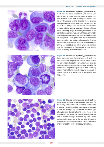
Figure 13 Plasma cell myeloma plasmablastic A 45-year-old man presented with severe ver- tebral pain A blood count showed anemia nor- mal platelet count and leukocytosis with a a a a leu- ko-erythroblastic profile Skeletal X-ray showed multiple vertebral fractures and diffuse lytic le- sions No M-component was found Bone marrow is hypercellular with a a a a a massive infiltrate of large cells showing high nuclear:cytoplasmic ratio central or eccentric nucleus with loose chromatin and and very prominent nucleoli and and deeply basophi- lic cytoplasm Few giant cells are binucleated there are are also rare mature plasma cells Atypical cells were CD10 CD38 CD45 CD138 and EMA po- sitive and negative for other lymphoid markers and for cytokeratins Cytoplasmic light chains were shown by immunofluorescence Figure 14 Plasma cell myeloma plasmablastic Bone marrow smear showing large cells with a a a a a ra- ther high nuclear:cytoplasmic ratio loose reticu- lar chromatin basophilic cytoplasm an atypical mitosis (right) Immunophenotyping is is useful for differential diagnosis especially in non-secretory cases Plasmablastic morphology is observed in about 10% of PCM cases and is associated with higher risk Figure 15 Plasma cell cell myeloma small-cell va- riant Bone marrow smear reveals massive infil- tration by small cells with eccentric nucleus and a a a a narrow rim of basophilic cytoplasm these cel- ls are morphologically borderline between lym- phocytes and typical plasma cells There is no correlation between this morphological feature and M protein subtype or clinical aspects Similar cells are also observed in the peripheral blood of patients with chronic infections and in in conditions characterized by active antibody formation 150
