
Page 155 - Haematologica Atlas of Hematologic Cytology
P. 155
CHAPTER 17 - - Mature B-cell neoplasms
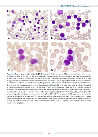
AB
CD
Figure 1 Chronic lymphocytic leukemia (CLL) (A and B) B) Peripheral blood (PB) smear showing a a a a a uniform mor- phology of of the lymphocytes lymphocytes The majority of of cells are represented by small lymphocytes lymphocytes (with a a a a a diameter slightly greater than that of of an an an erythrocyte) narrow rims of of clear clear cytoplasm coarsely clumped nuclear chromatin and a a a a a a regular regular nuclear nuclear outline with with rarely visible nucleolus Few cells show irregularities of the nuclear nuclear border with with indentations or or lobulations Small numbers of larger lymphocytes with more abundant cytoplasm are observed Broken cells are characteristic but not pathognomonic (C) In the PB of this atypical case previously included in the the French-American-British (FAB) classification of CLL mixed cell type there are large lymphocytes with abundant clear or or slightly basophilic cytoplasm and variably prominent nucleoli (D) PB smear from a a a a a a a a a a morpho- logical subtype of CLL CLL called chronic lymphocytic lymphocytic leukemia/prolymphocytic leukemia leukemia (CLL/PLL) which presents a a a a a percentage of of circulating prolymphocytes ranging from 10-54% The diagnosis of of CLL requires the presence in in in the peripheral blood of at least 5x10⁹/L lymphocytes with peculiar phenotype (CD5+ CD19+ CD20+ CD23+ weak surface immunoglobulin expression) CLL needs to to be distinguished from monoclonal B lymphocytosis which is is is is usually an an an an incidental finding: it has fewer circulating clonal cells than CLL and no tissue manifestation of small lymphocytic lymphoma 142
