
Page 106 - Haematologica Atlas of Hematologic Cytology
P. 106
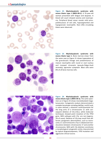
Figure Myelodysplastic syndrome with excess blasts type 1 1 (MDS 1) A 70-year-old woman presented with fatigue and dyspnea A blood cell count showed anemia and neutrope- nia Peripheral blood smear reveals mild aniso- poikilocytosis of red cells hyposegmented and hypogranular neutrophils Rare (3%) circulating blasts were observed Figure Myelodysplastic syndrome with excess blasts type 1 Bone marrow smear from the same case as as as Figure 21 shows hyperplasia of the granulocytic lineage and predominance of mature neutrophils with round or oval nucleus and clumped chromatin (pseudo-Pelger-Huët anomaly) agranular cytoplasm Blast cells were 8% of all bone marrow cells Figure 4 Myelodysplastic syndrome with excess blasts type 1 1 (MDS 1) The same speci- men as as in Figure 23 shows monolobulated mega- karyocytes Cytogenetic analysis demonstrated a a a a a a a a a a complex karyotype including del(5q) inv(12) and in in in in some some some mitoses a a a a a dicentric isochromosome for the long arms of of a a a a chromosome chromosome 17 17 17 with con- sequent deletion of of of 17p 17p (17p-) while molecular techniques showed point mutation of of the TP53 gene MDS subtypes with 17p- are are rare (approx 3% of of cases) Deletion of of 17p 17p may result from dif- ferent cytogenetic abnormalities such as as mono- somy 17 17 17 isochromosome 17q or or unbalanced tran- slocations between chromosome chromosome chromosome 17 17 17 17 and another chromosome chromosome MDS with 17p deletion constitutes a a a a a a a a morphological cytogenetic entity characterized by typical dysgranulopoiesis TP53 mutation and aggressive clinical course 93
