
Page 40 - 2020_01-Haematologica-web
P. 40
E. Mejia-Ramirez and M.C. Florian et al.
premature aging,111 including features of MDS.112 Trifunovic et al. concluded that there might be a causative link between mtDNA mutations in these mice and the aging phenotype.111 However, deep insights in the pheno- type and molecular signatures of aged HSC demonstrated that they are different from this ‘mutator’ strain, conclud- ing that aging is primarily independent of the accumula- tion of mitochondrial mutations.105 What is, indeed, observed is that aged HSC show deregulation that deteri- orates mitochondrial activity (reviewed by Oh et al.113).
Maintenance of mitochondrial homeostasis within the cell is also dependent on the regulation of nuclear mito- chondrial gene expression. For example, Mohrin et al. showed that SIRT7, a histone deacetylase, is normally located at the proximal promoters of ribosomal proteins and mitochondrial transcription factors within the nuclear genome. It also binds directly to NRF1, the master tran- scription factor of mitochondria. Eventually, SIRT7 leads to the inhibition of the expression of mitochondrial pro- teins in young HSC.114 Aged HSC have lower levels of SIRT7, thus allowing an increment in the expression of mitochondrial proteins. This, in turn, activates the PFSmt
(mitochondrial protein folding stress), a metabolic check- point that reduces HSC quiescence. Therefore, overex- pression of SIRT7 in aged murine HSC was shown to decrease the levels of mitochondrial protein expression associated with mitochondrial stress and to improve aged HSC function.114
Autophagy
Autophagy is a major regulator of mitochondria home- ostasis and a suppressor of the metabolism. It is regulated by nutrient-sensing pathways like mTOR or AMPK, which inhibit and activate autophagy, respectively. In gen- eral, autophagy decreases with aging, due to either the downregulation or the upregulation of critical autophagy proteins. In HSC, the age-related decrease in autophagy has been associated with lower capacity of glucose intake; mitochondrial autophagy would be attenuated in order to maintain necessary energy levels in old HSC and is rapidly induced by FoxO3 transcription factor when HSC are under metabolic stress.109
Genetic tools were instrumental in revealing the critical role of autophagy upon HSC aging.115 For example, in
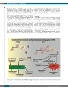
Figure 3. Metabolic homeostasis and proteostasis during aging in hematopoietic stem cells (HSC). Young HSC fine-tune several biological processes: they maintain a low metabolic rate, control protein degradation and regulate autophagy. Aged HSC show an unbalanced scenario where these processes lose their metabolic home- ostasis and proteostasis, converging into a status of metabolic and protein stress. Fasting is able to restore at least partially the activity of the UPRER and proteostasis in aged HSC. ROS: reactive oxygen species; UPR: unfolded protein response.
30
haematologica | 2020; 105(1)
