
Page 98 - 2019_11 Resto del Mondo-web
P. 98
R.T. Silver and S. Krichevsky
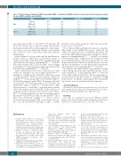
Table 2. Threshold values of hematocrit (HCT), hemoglobin (HGB), or red blood cell (RBC) count for men and women with associated area under the curve (AUC), specificity, and sensitivity.
Men
Women
Value
HCT (%) HGB (g/dL) RBC (x1012/L) HCT (%) HGB (g/dL) RBC (x1012/L)
Threshold
49.3 16.8 5.3 47.9 15.3 5.1
AUC
0.819 0.753 0.761 0.957 0.875 0.924
Specificity (%)
100.0 100.0 100.0 100.0 88.9 81.5
Sensitivity (%)
64.4 62.8 52.5 71.1 75.0 87.1
was measured in 225 of 410 (54.9%) PV patients. Of those that did not have a marrow biopsy, the majority had been encountered for only a single visit so that a mar- row biopsy was not temporally feasible or the patient was advised to have it performed with their primary hematologist.
In the absence of isotope studies and an initial marrow biopsy or SEV, it is important to evaluate the accuracy, as defined statistically, of the HCT, HGB, and RBC threshold values that are advocated to distinguish ETJAK2V617F from PV. We found overlap in HCT, HGB, and RBC values ranging from 25.0-54.7% indicating that a single red cell value will not effectively distinguish ETJAK2V617F from PV.
Such considerations have been overlooked in other studies. For example, it has been suggested that ETJAK2V617F patients are at higher risk for thrombosis than those with a CALR mutation.21 However, those patients diagnosed with ETJAK2V617F had a median SEV of 4.7 mU/mL (range: 0- 47 mU/mL) compared with CALR+ ET patients who had a median SEV of 9.4 mU/mL (range: 1.2-27 mU/mL). An unspecified number of ETJAK2V617F patients had a SEV below normal (i.e. <4 mU/mL)22 suggesting the possibility of PV. Since neither all red cell values, isotope studies, nor systematic marrow biopsies were reported, some of these ETJAK2V617F patients might have, in fact, had a higher risk of thrombotic events because they actually had PV.23 Thus, they were incorrectly assigned to a disease with a decreased expected survival.24 Of course, these issues do not occur in JAK2V617F wild-type, CALR+, or MPL+
patients because these mutations, with very rare excep- tions, do not occur in PV.18
It is of interest that our threshold values are coinciden- tally similar to the WHO 2016 criteria,6 which did not address the important topic of imperfect specificity and sensitivity. Although marrow biopsy and SEV are advan- tageous for distinguishing ETJAK2V617F from PV, it is unclear how frequently these examinations are being performed in actual clinical practice. Our data support their use even despite the discussed limitations. In the absence of resolv- ing these discrepancies, isotope RCM studies remain the gold standard for discriminating ETJAK2V617F from PV.
In summary, the clinical hematologist must be warned of the varying specificity and sensitivity and the consid- erable limitations of discriminating ETJAK2V617F from PV solely when using red cell values, and the importance of isotope, marrow, and SEV studies as outlined by WHO 2016 criteria.6 It remains undetermined how frequently any of these tests are performed in clinical practice.
Acknowledgments
We thank Dr. Paul Christos for statistical review. He was par- tially supported by the Clinical and Translational Science Center, Weill Cornell Medical College (UL1-TR000457-06).
Funding
This study was supported in part by the William and Judy Higgins Trust and the Johns Family Foundation of the Cancer Research and Treatment Fund Inc., New York, NY, USA.
References
1. Tefferi A, Thiele J, Orazi A, et al. Proposals and rationale for revision of the World Health Organization diagnostic criteria for polycythemia vera, essential thrombo- cythemia, and primary myelofibrosis: rec- ommendations from an ad hoc international expert panel. Blood. 2007;110(4):1092-1097.
2. Barbui T, Thiele J, Kvasnicka HM, Carobbio A, Vannucchi AM, Tefferi A. Essential thrombocythemia with high hemoglobin levels according to the revised WHO classification. Leukemia. 2014: 28(10):2092-2094.
3. Spivak JL. Myeloproliferative Neoplasms. N Engl J Med. 2017;377(9):895-896.
4. Silver RT, Chow W, Orazi A, Arles SP, Goldsmith SJ. Evaluation of WHO Criteria for Diagnosis of Polycythemia Vera: A
Prospective Analysis. Blood. 2013;
22(11):1881-1886.
5. Silver RT, Kiladjian JJ, Hasselbalch HC.
Interferon and the treatment of poly- cythemia vera, essential thrombocythemia and myelofibrosis. Exp Rev Hematol. 2013;6(1):49-58.
6. Arber DA, Orazi A, Hasserjian R, et al. The 2016 revision to the World Health Organization (WHO) classification of myeloid neoplasms and acute leukemia. Blood. 2016;127(20):2391-2405.
7. Barbui T, Thiele J, Gisslinger H, et al. Masked polycythemia vera (mPV): Results of an international study. Am J Hematol. 2014;89(1):52-54.
tion from essential thrombocythemia. Am J
Hematol. 2017;92(1):1062-1067.
9. Overhage JM, Ryan PB, Reich CG, Hartzema AG, Stang PE. Validation of a common data model for active safety sur- veillance research. J Am Med Inform Assoc.
2012;19(1):54-60.
10. Alvarez-Larran A, Ancochea A, Angona A,
et al. Red cell mass measurement in patients with clinically suspected diagnosis of polycythemia vera or essential thrombo- cythemia. Haematologica. 2012; 97(11): 1704-1707.
11. Johansson PL, Safai-Kutti S, Kutti J. An ele- vated haemoglobin concentration cannot be used as a surrogate marker for absolute erythrocytosis: a study of patients with polcythaemia vera and appartent poly- cythaemia. Br J Haematol. 2005;129(5):701-
8. Kvasnicka HM, Orazi A, Thiele J, Barosi G, Bueso-Ramos CE, Vannucchi AM.
European LeukemiaNet study on the repro-
ducibility of bone marrow features in 705.
masked polycythemia vera and differentia-
12. Silver RT, Gjoni S. The hematocrit value in
2204
haematologica | 2019; 104(11)
