
Page 52 - 2019_11 Resto del Mondo-web
P. 52
K.N. Ward et al.
to avoid unnecessary, potentially toxic, antiviral therapy. Chromosomally integrated human herpesvirus 6 should be suspected in the donor and/or recipient if HHV-6 DNA detection follows one of the patterns described in Table 3 or if HHV-6A is detected. Where necessary, CIHHV-6 can easily be excluded by a negative HHV-6 DNA test on a blood/serum sample taken pre-transplant from the recipi- ent or at any time from the donor. Individuals with CIHHV-6 have characteristic persistently high levels of HHV-6 DNA in whole blood (>5.5 log10 copies/mL) and in serum (100-fold lower than that in whole blood for a given patient).5,7 The level of DNA detected in plasma varies depending on the timing of separation from whole
blood.29
A ratio of one copy of HHV-6 DNA/cellular genome
confirms the diagnosis of CIHHV-6. Droplet digital PCR29 is the most accurate method as it gives an absolute num- ber. Comparison of two quantitative real-time PCR results (one for HHV-6 and one for a human gene present in all nucleated cells) is also acceptable albeit with a significant margin of error due to inherent assay imprecision.7 HHV- 6 DNA is present in hair follicles and nails exclusively in persons with CIHHV-6.4,19
• If CIHHV-6 is suspected, whole blood or serum or cel- lular samples or leftover DNA taken from donor and/or recipient pre-HSCT should be tested by quantitative PCR that distinguishes between HHV-6A and HHV-6B DNA. Testing plasma is not recommended.
• CIHHV-6 can be confirmed by evidence of one copy of viral DNA/cellular genome, or viral DNA in hair folli- cles/nails, or by FISH demonstrating HHV-6 integrated into a human chromosome.
Tests for chromosomally integrated human herpesvirus 6 reactivation
This must be confirmed by virus culture plus viral genome sequencing to confirm identity of the viral isolate with the integrated virus.
Human herpesvirus 6B end-organ disease and other outcomes post-hematopoietic stem cell transplantation
Human herpesvirus 6B primary infection versus reactivation
Only two cases of primary HHV-6B infection after allo- geneic HSCT have been reported; these were in very young children and were accompanied by fever and rash.30,31 In contrast, various end-organ diseases and other complications post-HSCT have been associated with HHV-6B reactivation. But apart from encephalitis and fever with rash, the evidence for causation is moderate or weak (Table 4).
Human herpesvirus 6B encephalitis and its definition
The first described encephalitis case32 was followed by many confirmatory reports.33 Zerr and Ogata analyzed the accumulated published data and provided evidence for a causal association between HHV-6 and encephalitis using the Bradford Hill criteria.34
The most frequent cause of encephalitis after allogeneic transplant is HHV-6. When the species is identified, it is
almost invariably HHV-6B. Of the only three reported patients with HHV-6A encephalitis, one had an atypical presentation and the other two had unrecognized CIHHV-6.9 In one of these two, testing of archived sam- ples confirmed CIHHV-6A pre-HSCT,35 but the question remained as to whether reactivation of the virus causing encephalitis or an alternative unidentified cause was responsible. Whether CIHHV-6B can reactivate causing encephalitis is theoretically possible, but requires viral culture and sequencing to distinguish childhood-acquired HHV-6B from integrated virus.
Human herpesvirus 6B encephalitis typically presents early as post-transplant acute limbic encephalitis (PALE). CSF protein and cell counts are often unremarkable (see Table 5 for further clinical features). Although magnetic resonance imaging (MRI) may be negative at the start of the disease, changes in the temporal lobe are demonstrat- ed in approximately 60% of cases.36 However, similar observations occur in limbic encephalitis caused by other infectious agents.37 Extrahippocampal abnormalities may
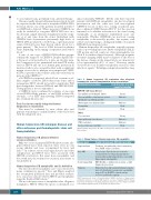
Table 4. Human herpesvirus 6B reactivation after allogeneic hematopoietic stem cell transplantation: disease associations.
Epidemiological associations
HHV-6B end-organ disease
Encephalitis (predominantly limbic)
Non-encephalitic central nervous system
dysfunction e.g. delirium, myelitis Myelosuppression, allograft failure Pneumonitis
Hepatitis
Other
Fever and rash
Acute graft-versus-host disease
CMV reactivation
Increased all-cause mortality
Level of in vitro or in vivo support for causation
Strong
Moderate
Moderate Weak Weak
Strong
Moderate
Moderate
Weak
HHV-6B: human herpesvirus 6B; CMV: cytomegalovirus. Adapted from Table 29.2 in Hill and Zerr.98
Table 5. Clinical features of human herpesvirus 6B encephalitis.
Disease onset
Symptoms/signs
Brain MRIa
Cerebrospinal fluid
Prognosis
Usually 2-6 weeks post HSCT, but can be later
Confusion, encephalopathy, short-term memory loss, SIADH, seizures, insomnia
Often normal. Typically but not exclusively, circumscribed, non-enhancing, hyperintense lesions in the medial temporal lobes (especially hippocampus and amygdala)
HHV-6B DNA, +/- mild protein elevation, +/-mild lymphocytic pleocytosis
Memory defects and neuropsychological sequelae in 20-60%. Death due to progressive encephalitis in up to 25% of all HSCT recipients and up
to 50% of cord blood recipients
HSCT: hematopoietic stem cell transplantation; SIADH: syndrome of inappropriate antidiuretic hormone secretion; MRI: magnetic resonance imaging; HHV-6B: human herpesvirus 6B. aFeatures of T2, fluid attenuation, inversion recovery (FLAIR) and diffu- sion weighted-imaging sequences. Modified from Hill and Zerr.99
2158
haematologica | 2019; 104(11)
