
Page 204 - 2019_11 Resto del Mondo-web
P. 204
B. Brenig et al.
Supplementary Table S1). These males had increased aPTT ratios of 2.93 (#53) to 4.76 (#51) indicative of defects in the intrinsic coagulation pathway and also reduced FIX con- centrations in the blood as is normally the case in hemo- philia B. The affected dog #3 presented only 2% of the standard FIX concentration. The female carriers #4 and #6 showed aPTT ratios within the reference range. FIX con- centrations, however, were slightly below the reference range (#6). This was not surprising as it has been shown that minute reductions in FIX concentrations might not always be reflected in an aPTT increase because of the sensitivity of commercial reagents.43 The clinical signs together with the blood coagulation parameters and X- linked transmission supported the diagnosis of hemophil- ia B. The definite clinical diagnosis prompted us to search for the molecular cause initially on the DNA level. The canine F9 gene is located on chromosome X (CFAX) between positions 109,501,341 (transcription start site) and 109,533,798 and has a length of 32,458 bp (NC_006621.3, CanFam3.1). The canine F9 gene, similarly to that of other mammals, has eight exons with an open reading frame of 1,356 bp coding for 452 amino acids.44 DNA of female carriers #4 and #6 was subjected to whole genome sequencing and aligned to the canine reference F9 gene sequence. Surprisingly, only six sequence variants outside the coding regions of F9 were identified (Table 1). Five variants were located in introns and were excluded as the cause of the hemophilia B in the Hovawarts because these variants were also detected in unaffected controls. The remaining variant (deletion) was located in the pro- moter of F9 73 bp upstream of the start codon (Online Supplementary Figures S1 and S3). As this deletion was located within a putative transcription factor binding site of HNF4α and AR, which had been shown in humans to be important for F9 expression and mutated in hemophilia B Leyden and Brandenburg,31,32 this position was analyzed in more detail.
Figure 1 shows the segregation of the nucleotide deletion in the affected Hovawart family. The female carriers #4 and #6 were heterozygous, as evidenced by the overlap-
ping peaks with similar heights 5’ of the deletion position. The affected male #3 was hemizygous for the deleted allele whereas his sister #5 and cousin #7 were homozy- gous wildtype. Genotyping of 1,298 dogs (including 83 dif- ferent breeds, 720 unrelated Hovawarts, and 12 Hovawart family members) demonstrated the occurrence of the dele- tion only among members of the affected Hovawart family (Table 2, Online Supplementary Table S2). To provide proof that the deletion represented the causative genetic variant and resulted in the low expression of F9, functional analy- ses using electrophoretic mobility shift and luciferase reporter assays were performed.
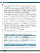
Table 1. DNA sequence variants in the canine F9 gene determined by next-generation sequencing of DNA of animals #4 and #6.
As shown in Figure 2, no binding of recombinant HNF4α to the mutated promoter region was detected. On the other hand, the AR lysate clearly showed binding to both fragments and hence the deletion seems not to influence AR binding to the androgen-responsive element in the canine F9 promoter. To analyze the effect of the promoter variant on F9 expression, wildtype and mutated promoter fragment luciferase constructs were transfected into Hep G2 cells. As shown in Figure 3 the mutated promoter frag- ment resulted in a statistically highly significant (P=2.2x10- 6) reduction of the relative response ratio to approximately 34.6% of the wildtype promoter in the presence of C/EBP (+ C/EBP). C/EBP is clearly also an important transcription factor in the regulation of the canine F9 promoter as shown when C/EBP was not co-transfected (- C/EBP). In the absence of C/EBP the relative response ratio of the wild- type promoter was 29.8%. On the other hand there were no significant differences between the mutated promoter fragment (+/- C/EBP) and the wildtype promoter fragment (- C/EBP). As for variants of the HNF4α site, disruption of the C/EBP binding site has also been shown to be causative for hemophilia B in humans.45,46
Discussion
As in humans, hemophilia A and B are also rare diseases in dogs caused by sequence variants in the coagulation
Position Ref/Alta
X:109501492 C/-
X:109504229 C/-
X:109505462 -/AG
X:109507446 -/A
X:109510986 G/A
X:109524055 A/G
Gene region
5’-flanking region
intron 1
intron 1
intron 2
intron 3
intron 6
HGVSb g.
NC_006621.3:g.109501492delC
NC_006621.3:g.109504229delC NC_006621.3:109505462_109505463insAG NC_006621.3:109507446_109507446insA NC_006621.3:g.109510986G>A NC_006621.3:g.109524055A>G
aRef/Alt: reference nucleotide/alternate nucleotide; bHGVS: Human Genome Variation Society (http://www.hgvs.org).
Table 2. F9 genotype frequencies
Genotype
C/C
C/-
-/-
HBa affected (n=1)
1
Hovawart
HB carrier (n=2) Control, related (n=12)
Control, unknown relationship (n= 720)
720
Other breedsb Controls (n= 567)
567
2
12
aHB: hemophilia B; bdog breeds used as controls are listed in Online Supplementary Table S2.
2310
haematologica | 2019; 104(11)
