
Page 111 - 2019_11 Resto del Mondo-web
P. 111
K-Ras mutation depletes pre-leukemic HSC
between samples was determined using an analysis of variance (ANOVA) for multiple comparisons following Tukey’s multiple comparisons test.
Results
Aml1-ETO ameliorates key features of the myeloproliferative neoplasm phenotype caused by K-RasG12D
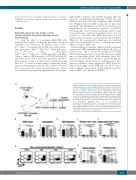
To study the effect of combining Aml1-ETO with mutant K-Ras, whilst avoiding the previously described spontaneous recombination in primary KrasG12D/+;Mx1- Cretg/+ mice,19,20 we generated E14.5 FL cells with the neces- sary genotypes: Aml1ETO/+;Mx1-Cretg/+ (AM genotype); KrasG12D/+;Mx1-Cretg/+ (KM genotype); Aml1ETO/+;KrasG12D/+;Mx1-Cretg/+ (AKM genotype); and Mx1- Cretg/+ controls (CON genotype); all CD45.2 allotype. FL cells were used as these have been previously shown to have either no or minor spontaneous recombination using Mx1-Cre.29 To create a scenario where competitive advan- tage and disadvantage of mutant HSC could be observed, mice were generated where the hematopoietic system was partially repopulated by experimental cells and par-
A
tially by WT competitor cells (CD45.1 allotype). This was done by competitively transplanting 2.5x10⁵ FL cells (CD45.2) and 1x10⁶ WT BM competitor cells (CD45.1) into lethally irradiated CD45.1 recipients, as previously described.27 Recombination was induced four weeks post- transplantation by subcutaneous injection of poly(I:C), with all groups of mice treated, including controls. Long- term monitoring of primary transplanted mice was not possible as both KM and AKM developed a T-cell leukemia (data not shown), as previously reported for KM.20 Therefore, the hematopoietic phenotype was analyzed eight weeks post-poly(I:C) before T-cell leukemia devel- opment occurred (Figure 1A).
AM-transplanted recipients displayed mild leukopenia compared to CON-transplanted mice (Figure 1B), due to a decrease in B and T cells in the peripheral blood (PB) (Online Supplementary Figure S1A and B), whereas no differ- ence in hemoglobin levels, BM cellularity, Mac1+Gr1lo myeloid cells in the PB or spleen, or spleen weight was observed (Figure 1C-H). However, AM showed a 27% decrease in platelet count compared to CON (Figure 1I). These results suggest Aml1-ETO affects B and T cell, as well as platelet development, in line with known func- tions of AML1 (also known as RUNX1).30,31
Figure 1. Aml1-ETO ameliorates the key features of the myeloproliferative neoplasm phenotype caused by K-RasG12D. (A) Schematic representation of in vivo competitive transplant experiment. (B-I) Analysis of recipients of Mx1-Cretg/+ controls; CON (n=13), Aml1ETO/+;Mx1-Cretg/+; AM (n=14), KrasG12D/+;Mx1-Cretg/+; KM (n=12) and Aml1ETO/+;KrasG12D/+;Mx1-Cretg/+; AKM fetal liver (FL) (n=14) for (B) peripheral blood (PB) white blood cell (WBC) count. (C) PB hemoglobin levels. (D) Bone marrow (BM) cellularity per tibia and femur. (E and F) CD45.2 Mac1+Gr1lo myeloid cells as absolute number in the PB (E) and spleen (F). (G) Representative FACS plots of Mac1+Gr1+
and Mac1+Gr1lo myeloid cells as LiveCD19–CD4–CD8a–NK1.1–CD45.2+ cells across all experiments in the PB. (H) Spleen weight. (I) Platelet count. Results were generated in three independent experiments. The results were analyzed using multiple com- parison ANOVA and are presented as mean±Standard Deviation. *P<0.05; **P<0.01; ***P<0.001; ****P<0.0001.
a percentage of
BCD
G
EF
HI
haematologica | 2019; 104(11)
2217
