
Page 83 - 2019_10 resto del Mondo_web
P. 83
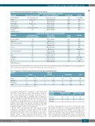
Immunosuppressive therapy for pediatric aplastic anemia
Table 6. Clonal cytogenetic abnormalities at diagnosis and at follow up.
Acquired chromosomal abnormalities at diagnosis
Abnormality
del(13)(q12q21)*
Status at follow up evaluation
Decreased clone size
Best response to initial therapy
Relapse at two years
CR by 12 months NR by 3 months NR by 6 months CR by 3 months CR by 6 months NR by 12 months
Second therapy
IST
None None BMT None None IST
Second therapy
None
None IST None None BMT BMT None IST IST IST IST
Outcome
Death, MVA
Alive Alive Alive Alive Alive Alive
Outcome
Alive
Alive Alive Alive Alive Death, PNA Alive
Alive
Alive Death, ALL Death, AML Alive
del(7q)
add(11)(q23)*
del(16)(q22)*
add(14)(q11.2)*
del(13)(q12q14)*
+mar[2]* N.A. *hATG/CyA subjects
Not detected N.A.
Not Detected N.A. Decreased clone size
Post-treatment acquisition of chromosomal abnormalities
Abnormality
del(13q)(q14q22)*
i(X)(p10)* der(5)t(1;5)(q11;q11.2)* -7*
+14,-18*
-7 -5,-7,del(7q),del(20p)* 8*
i(2q)*
-7,del(7q)*
del(7q22)*
-7*
*hATG/CyA subjects
Time from diagnosis
to first detection (months)
4.6
5.5 66.5 37.1 4.5 20 4.3 71 67.7 30.3 67 8
Best response to initial therapy
CR by 6 months
CR by 3 years NR by 12 months VGPR by 2 years CR by 12 months NR by 2 years NR by 6 months VGPR by 5 years Relapse at 2 years NR by 12 months Relapse at 2 years NR by 6 months
NR: no-response; N.A.: not available; VGPR: very good partial response; CR: complete response; IST: immunosuppressive therapy; BMT: bone marrow transplant; MVA: motor vehicle accident; PNA: pneumonia; ALL: acute lymphoblastic leukemia; AML: acute myeloid leukemia.
Table 7. Comparison of baseline cytogenetic clones with subsequent clonal abnormalities.
Baseline* Follow Up
Normal Abnormal Not Evaluable
N%N%N%
Normal 137 51.9 12 4.6 115 43.6
Abnormal 3 42.9 2 28.6 2 28.6
Not Evaluable 16 37.2 1 2.3 26 60.5
Total 156 49.7 15 4.8 143 45.5
Total
264
7 43 314
*Cytogenetics or fluorescence in situ hybridization at diagnosis.
assessments performed at diagnosis, seven (3%) had detectable clonal chromosomal abnormalities (Table 6). Six of these patients had follow-up cytogenetic assess- ments. Two patients had a del(13q) clone at diagnosis, which remained detectable through the duration of follow up (range: 33-49 months) but was not associated with acquisition of additional chromosomal abnormalities. In contrast, other small clones present at diagnosis, including del(7q) in one patient and del(16q) in one patient, were no longer detectable at follow-up assessment.
Of the 171 total patients who had follow-up BM metaphase cytogenetics (n=160) and/or FISH (n=109) assessment performed after IST initiation, 12 (7.0%) patients had evidence of new clonal chromosomal abnor- malities (Table 7). One additional subject had a clonal abnormality [+der(14;21)(q10;q10)] at 143 months from
Table 8. Subsequent treatments. Subsequent All patients (N=314)
hATG/CyA (N=264) Treatment N % N %
2nd therapy 110 35 92 35
3rdtherapy 35 11 33 13
4th therapy 3 1 3 1
the time of initial diagnosis; however, the baseline status was unknown. The most common genetic alteration after IST was loss of chromosome 7 [either -7 or del (7q)] occur- ring in six patients (3.5%), all of whom had normal cyto- genetics at baseline. Three patients had a del(13q) clone detected during follow-up BM assessments of which one was acquired after treatment. Among those patients with
haematologica | 2019; 104(10)
1979
