
Page 69 - 2019_10 resto del Mondo_web
P. 69
FANCP (SLX4)
FANCP (SLX4)
FANCP (SLX4)
FANCQ (ERCC4)
FANCR (RAD51)
FANCS (BRCA1)
FANCS (BRCA1)
FANCS (BRCA1)
FANCS (BRCA1)
FANCS (BRCA1)
FANCT (UBE2T)
FANCU (XRCC2)
FANCW (RFWD3)
FANCW (RFWD3)
FANCW (RFWD3)
FANCW (RFWD3)
chr16:3640407 CAGCTGG/C
chr16:3639742 CCT/C chr16:3639379 T/TG chr16:14042182 C/G chr15:40994106 C/T chr17:41258497 A/T chr17:41245553 G/GAAA chr17:41244748 G/A chr17:41244333 AG/A chr17:41226421 C/CA chr1:202304773 C/T chr7:152346394 TA/T chr16:74695317 G/A chr16:74685992 G/GA chr16:74678352 C/T chr16:74660405 G/A
c.3226_3231del
c.3895_3896del c.4259_4260insC c.2729C>G c.328C>T c.188T>A c.1995_1997insTTT c.2659C>T c.3214delC c.4664_4665insT c.109+1G>A c.175delT
c.31C>T
c.546_547insT
c.988-1G>A
c.2017C>T
p.P1076_A1077del
p.R1299fs p.I1421fs p.S910X
p.R110X
p.L63X p.N665_L666insF p.Q887X p.L1072fs p.E1556fs aberrant splicing p.T59fs
p.Q11X
p.Q183fs
aberrant splicing
p.R673X
0.0001
0.0001
0.0001
0.0001
0.0003
0.0003
0.0001
0.0001
0.0001
0.0001
0.0004
0.0001
0.0001
0.0001
0.0001
0.0001
Molecular diagnosis and clinical features of 117 Japanese FA patients
continued from previous page
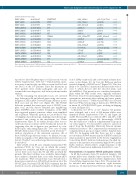
These data were obtained from 3.5KJPNv2 site mutations. Missense mutation variants
database (https://jmorp.megabank.tohoku.ac.jp/201811/). *We focused on nonsense mutations, frameshift mutations, and splicing were not included.
reported to affect FA phenotypes (see Discussion section) (Online Supplementary Table S2).5,10 Unfortunately, muta- tions were found in only one allele in seven (six FA-A and one FA-G) of the 107 patients. Since the mutations in these patients were clearly pathogenic and rare, we assumed this was diagnostic, and did not perform further analysis.
For the remaining ten unclassified cases, we screened large deletions in FA and related genes using our custom- designed aCGH in 2014. It revealed large deletions in two FA-B cases and one FA-T case (Figure 1B). The FANCB deletions spanned the entire genic area of FANCB (com- plete loss), and the defects extended into neighboring genes MOSPD2 and/or GLRA2. Reanalysis of the WES data suggested putative junctions, where were amplified and sequenced. While the junction in Case 60 had a 3 bp overlapping microhomology, implying microhomology- mediated end joining as the mechanism (see Online Supplementary Figure S1 for further details), there was no such homologous sequence in the break point in Case 61, suggesting that the re-ligation was mediated by non- homologous end joining (Figure 1B).15 Two cases of entire FANCB deletion have been described in the literature16,17 without elucidation of the junctional sequence. All of these FANCB large deletions seem to be distinct, but uni- formly accompany severe phenotypic malformations (see below). The FA-T case with a large deletion was previous- ly described.6
After aCGH, seven FA cases remained unclassified. We performed WGS for Case 64, in which the parents’ genome was available, and RNA-seq analysis was carried out for three cases (Cases 62, 98, and 104), in which the patients' fibroblast cell lines were available. Interestingly, these analyses identified three cases with aberrant splice site mutations. WGS revealed that Case 64 harbored a homozygous mutation (c.1154+5G>A) in intron 12 of the FANCC gene. Real-time PCR (RT-PCR) confirmed that the mutation caused a splicing abnormality, resulting in reten-
tion of 120bp of intron 12 and a subsequent in-frame non- sense codon (Figure 1C). In Case 62, RNA-seq analysis revealed skipping of FANCB exon 7 (Figure 1D). This was likely to be caused by a mutation in the first nucleotide of exon 7, which did not alter the encoded amino acid (p.Leu499Leu). This mutation was considered non-patho- genic when the WES results were originally evaluated. However, it has been increasingly recognized that similar synonymous mutations affect splicing and cause genetic disorders and cancer.18,19 RNA-seq and WES also revealed that Case 98 had a homozygous mutation (c.3350+5G>A) in intron 12 of PALB2/FANCN gene, resulting in skipping of exon 12 (Figure 1D).
Collectively, 113 (97%) of 117 Japanese FA patients were subtyped, and a total of 215 mutant alleles were identified (Online Supplementary Table S2 and Figure 2A and B). FA-A and FA-G accounted for 58% and 25% of FA patients, respectively (Figure 2A). Interestingly, FANCB was the third most common complementation group in our series (approx. 3%). In notable contrast to a previous report from the Rockefeller University Fanconi Anemia Mutation Database,20 FA-C represented an extremely rare complementation group in Japan (Online Supplementary Table S1). In keeping with this, there was not a single record with an IVS4+4 mutation in the 3.5KJPN or the East Asian population represented in the Exome Aggregation Consortium (ExAC) database. In Europeans, the allele fre- quency of the mutation was relatively high (0.04%) in the ExAC database, which reflects a high frequency of the IVS4+4A>T mutation in Ashkenazi-Jewish FA-C cases.21
Characteristics of Japanese FANCA pathogenic variants In 68 FA-A patients (from 59 unrelated families), 130 mutant alleles were identified that consisted of 55 differ- ent FANCA variants (listed in Online Supplementary Table S3 and Online Supplementary Figure S3A). The mutant alle- les included nine missense mutations, eight nonsense mutations, 16 small insertions/deletions (indels), 12 large
haematologica | 2019; 104(10)
1965
