
Page 46 - 2019_10 resto del Mondo_web
P. 46
P. Valent et al.
with repeated investigations of all disease-related parame- ters. A summary of non-clonal and clonal conditions potentially preceding CMML is shown in Table 4. With regard to criteria delineating non-clonal pre-diagnostic conditions, like idiopathic cytopenia of undetermined sig- nificance from the clonal conditions described above (CHIP, CCUS, CHOP), we refer the reader to the pertinent literature.69,71,73
Peripheral blood and bone marrow smears: proposed standards and recommendations
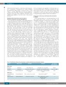
As in other myeloid neoplasms, a thorough examination of appropriately prepared and stained BM and PB smears is a crucial diagnostic approach in suspected CMML. It is standard to examine and count at least 100 leukocytes in the PB film and 200-500 nucleated cells in well-prepared thin BM films. BM cellularity, the erythroid-to-myeloid (E:M) ratio, and the percentage of blast cells (including monoblasts and promonocytes), monocytes, mast cells, and other myeloid cells must be recorded (reported) in each case. As in patients with MDS, at least 10% of cells in one of the major BM lineages (erythroid and/or neu- trophilic and/or megakaryocytic) must be dysplastic to meet the dysplasia criterion for CMML.13-18 It is also stan- dard to study well-prepared and appropriately stained PB smears in CMML and to report the percentage of circulat- ing monocytes, including normal (mature) and abnormal (immature) monocytes, blast cells, other immature myeloid cells, dysplastic (hypogranulated) neutrophils and other cell types in the PB. Overall, the same standards and recommendations that count for the evaluation of MDS by morphology (BM and PB stains)12,80-83 also apply in cases with (suspected) CMML.13-18 An important point is the clas- sification of blast cells and monocytic cells in CMML (Table 5).16,84 Blast cell types detectable in CMML include myeloblasts, monoblasts and also promonocytes (even if not named blast cells) (Table 5). Monocytes should be clas- sified as normal (mature) or abnormal (immature).16,84 The morphological criteria used to distinguish between these cell types are presented in Table 5. Together with morphol- ogy, cytochemical staining for non-specific esterase can also assist in the cytological delineation between mono- cytes, monoblasts and promononcytes.16 An important aspect is that in many patients, megakaryocyte dysplasia is
better documented and quantified in BM histology sec- tions than in BM smears. Therefore, megakaryocyte dys- plasia should only be recorded in BM smears when a suffi- cient number of these cells can be detected. Finally, the morphology of mast cells, when detected, should always be reported using established criteria and standards.85
Bone marrow histology and immunohistochemistry in CMML
A thorough investigation of an appropriately processed and stained BM biopsy section by histology and immunhistochemistry is standard in all cases with known or suspected CMML or a suspected pre-CMML condi- tion.14-16,30,86 Notably, BM histology and immunhistochem- istry are essential approaches to confirm the diagnosis of CMML and to exclude AML and other CMML-mimick- ers. Moreover, BM histology and immunhistochemistry may provide important additional information, including that on BM fibrosis, focal accumulations of blast cells, increased angiogenesis, atypical (dysplastic) megakary- ocytes, a hypocellular BM or concomitant mastocytosis (Online Supplementary Table S4).33-35,86 The evaluation and enumeration of CD14+ monocytes, CD34+ progenitor cells and CD117+/KIT+ cells (progenitors and mast cells) by immunhistochemistry in BM biopsy sections repre- sent an integral part of the diagnostic assessment. These approaches can also prevent diagnostic errors. For exam- ple, when the smear is of suboptimal quality, a prelimi- nary diagnosis of CMML may change to AML based on BM histology and CD34 immunhistochemistry.
BM biopsy specimens are usually taken from the iliac crest and should be of adequate length (≥2 cm). The spec- imen should be fixed in neutral formalin (or alternative standard fixation), decalcified in EDTA (for at least 8 h) or by alternative standard decalcification, and embedded in paraffin-wax. Ideally 2-3 μm thin sections should be pre- pared. Routine stains include hematoxylin-eosin, Giemsa, Prussian blue, AS-D chloroacetate esterase, toluidine blue and silver impregnation (Gömöri’s stain). BM cellularity should be measured and reported according to published standards.87,88 For routine purposes, the pathologist should determine the cellularity as ‘normocellular’, ‘hypocellu- lar’, or ‘hypercellular’, based on an age-adapted esti- mate.89 The presence of variable degrees of BM fibrosis
Table 5. Classification of blast cells and monocytes in patients with chronic myelomonocytic leukemia.
Cell type
Blast cells:
Myeloblast
Monoblast Promonocyte
Monocytes:
Abnormal/immature monocyte
Mature monocyte
Nuclear shape
Round/oval
Round/oval Convoluted/indented*
Convoluted/indented
Lobulated/ indented
Chromatin
Fine with nucleoli
Delicate/lace-like, nucleoli Delicate/lace-like,
nucleoli
More condensed, rare nucleoli Condensed, no nucleoli
Cytoplasm
Basophilic, rare or no granules
Basophilic, rare azurophilic granules Variably basophilic, variably azurophilic
granules
Intermediate basophilic**
Gray or pinkish with occasional azurophilic granules
and vacuoles
Size relative to mature monocytes
Smaller
=
Smaller
Large (20-30 μM) Large
*The most important feature discriminating promonocytes from monoblasts. **Less basophilic than promonocytes and more basophilic than mature monocytes.
1942
haematologica | 2019; 104(10)
