
Page 42 - 2019_10 resto del Mondo_web
P. 42
P. Valent et al.
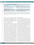
Table 2. Overview of special variants of chronic myelomonocytic leukemia.
Special variant
Oligomonocytic CMML
SM with concomitant CMML = SM-CMML
CMML with a concomitant myeloid neoplasm* expressing a classical MPN- driver, such as JAK2 V617F, BCR-ABL1
or rearranged PDGFRA/B*** or FGFR1.
CMML with expression of a molecular MPN-driver – examples: CMML with JAK2 V617F or CMML with a rearranged PDGFRA/B or CMML with rearranged FGFR1.
CMML with a concomitant lymphoid/lymphoproliferative neoplasm
Key diagnostic features that discriminate the variant from classical CMML
Absolute PB monocyte count <1x10⁹/L
WHO criteria for SM fulfilled; in most patients CMML monocytes exhibit
KIT D816V
WHO criteria for a classical MPN, such as CML**, PMF, or a myeloid neoplasm
with rearranged PDGFRA/B are fulfilled in addition to the criteria for CMML.
Molecular drivers of classical MPN, such as JAK2 V617F**** or rearranged PDGFRA/B*** are found but diagnostic criteria for such classical MPN are not fulfilled (only criteria for CMML are met)
WHO criteria for a lymphoid neoplasm are fulfilled
*These conditions must be separated from MPN with concomitant monocytosis that do not fulfil the diagnostic criteria for CMML.**Unlike in SM-CMML, in which monocytes display KIT D816V or CMML with rearranged PDGFRA, the CMML monocytes must not express BCR-ABL1 in patients with CML plus CMML. ***Several different translocations and fusion genes involving PDGFRA or PDGFRB may be detected, such as the t(5;12) associated with the TEL-PDGFRB fusion gene. ****JAK2 V617F itself counts as a feature of MPN; therefore, detection of JAK2 V617F can confirm the diagnosis of CMML (as MPN/MDS overlap disease) when other signs of myeloproliferation are absent (e.g., no splenomegaly and no leukocytosis).CMML:chronic myelomonocytic leukemia;PB:peripheral blood;SM:systemic mastocytosis;WHO:World Health Organization;MPN:myelo- proliferative neoplasm; CML: chronic myeloid leukemia; PMF: primary myelofibrosis.
We therefore recommend that in each case, deeper (full) prognostication should follow using multiparametric scor- ing systems (see later). It should be noted, however, that grading of CMML has only been validated in the classical form of CMML, not in special CMML variants. Therefore, although grading is also recommended for special CMML entities, it is not regarded standard and the result must be interpreted with caution in these patients.
Special variants of CMML: overview
As mentioned before, the classical form of CMML meets all pre-requisite criteria, and no signs (including molecular features) of an additional, concomitant BM neoplasm are detected. The special variants of CMML form a heterogeneous group of neoplasms comprising distinct clinical and biological entities. In one group of patients, the relative monocyte count (≥10%) is fulfilled without resulting in an absolute count ≥1×10⁹/L, preclud- ing the diagnosis of ‘classical CMML’. Most of these patients are diagnosed as having MDS or MPN/MDS- unclassified by WHO criteria. In another group of patients, a molecular signature suggestive of a different type of myeloid neoplasm is detected but only the criteria for CMML (not those for the other neoplasm) are met. Such an example is CMML with JAK2 V617F (without definitive evidence of a concomitant MPN). In a third group, CMML co-exists with another BM neoplasm, such as MPN or mastocytosis. In these patients, additional blood count abnormalities (e.g., eosinophilia), an elevated serum tryptase level and/or BM fibrosis, may be detected.
All variants of CMML (classical and special) can occur as a primary CMML or as a secondary CMML following a ‘mutagenic’ event, such as chemotherapy (therapy- related CMML). In addition, our faculty is of the opinion, that the term secondary CMML may also be appropriate for those patients who develop CMML (months or years) after another indolent myeloid neoplasm, such as a MDS or systemic (indolent or aggressive) mastocytosis, had been diagnosed. In the following paragraphs, the clinical features and diagnostic criteria of special (atypical) vari- ants of CMML are proposed and discussed. An overview of the special variants of CMML is provided in Table 2.
Oligomonocytic CMML
Over the past few years, more and more cases of cytopenic patients exhibiting relative monocytosis (≥10%) and moderately increased absolute blood mono- cytes not reaching the required threshold to diagnose classical CMML (1.0x10⁹/L) have been described. These cases have recently been referred to as oligomonocytic CMML.30 According to the WHO classification most of these patients would be classified as having MDS (with monocytosis) or perhaps MPN/MDS-unclassifiable. However, most of these patients exhibit typical features of CMML, including a typical morphology of PB and BM cells, splenomegaly, and CMML-related molecular fea- tures (e.g. mutations in TET2 and SRSF2).30-32 Some of these patients have prominent BM monocytosis without diagnostic PB monocytosis at diagnosis.30,32
Whereas several of these cases remain stable without progression, the majority will develop ‘overt’ CMML or, eventually, secondary AML during follow-up. Therefore, oligomonocytic CMML may also be regarded as a poten- tial pre-phase of classical CMML. Our faculty is of the opinion that the term oligomonocytic CMML should be used in clinical practice. Diagnostic pre-requisite criteria for oligomonocytic CMML are: (i) persistent (lasting at least 3 months) absolute peripheral monocytosis of 0.5- 0.9×10⁹/L and relative blood monocytosis (≥10% of blood leukocytes); (ii) exclusion of BCR-ABL1+ leukemia, classi- cal MPN and all other myeloid neoplasms that can explain monocytosis; and (iii) a blast cell count of 0-19% in PB and/or BM smears and exclusion of all histopatho- logical, morphological, phenotypic, molecular and cyto- genetic signs that count as proof of AML. Diagnostic dys- plasia in one or more of the three major BM lineages (≥10%) must also be documented. If dysplasia is lacking or ‘sub-diagnostic’ (<10%), the presence of cytogenetic or molecular lesions (mutations) typically found in CMML and/or the presence of CMML-related flow cytometry abnormalities, may also lead to the conclusion that the patient has oligomonocytic CMML provided that the other diagnostic criteria described above are fulfilled and all other myeloid neoplasms have been excluded. The proposed criteria for oligomonocytic CMML are listed in
1938
haematologica | 2019; 104(10)
