
Page 12 - 2019_10 resto del Mondo_web
P. 12
Editorials
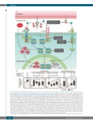
Figure 1. AXL promotes acute myeloid leukemia (AML) progression upon development of resistance. (A) Stromal cells were shown to express cytokines and ligands (see 1 and 2) (such as IL3 or FLT3-L, solid arrow) to a greater extent than AML cells (dashed arrow). Additionally, stromal cells were shown to produce GAS6 (see 3), which activates AXL receptor signaling. JAK2 binds to the cytosolic juxta-membrane region of dimeric cytokine receptors such as IL3R via the BOX1 and BOX2 receptor motifs upon stimulation by respective cytokines (see 2). JAK2 activation triggers STAT5 signaling, which promotes oncogenic gene transcription to propagate cancer cell survival, proliferation, and metabolic reprogramming. Additionally, STAT5 was shown to bind to the AXL promoter to induce expression. Mutated FLT3 (FLT3-ITD, yellow stars), frequently found in AML patients, can induce phosphorylation of STAT5 directly. Therapeutic agents used by Dumas et al.2 to target these key proteins/pathways in AML are summarized in gray boxes. Hypoxia induced by the stromal microenvironment induces expression of HIF transcription factors, and STAT5 can induce expression of HIF2α. Subsequently, HIF can bind to HRE elements in the AXL promoter. AML: acute myeloid leukemia; IL3: Interleukin 3; IL3R: Interleukin 3 receptor; GM-CSF: granulocyte-macrophage colony-stimulating factor; TPO: thrombopoietin; FLT3: Fms-like tyrosine kinase 3; ITD: internal tandem dupli- cation; FLT3-L: Fms-like tyrosine kinase 3 ligand; GAS6: growth arrest-specific 6; HIF: hypoxia-inducible factor; HRE: hypoxia-response element; JAK: Janus kinase; STAT: signal transducer and activator of transcription. (B) AXL mRNA expression levels in patients with hematopoietic cancers using data from the Oncomine data- base. Box plots showing human hematopoietic cancers with significant upregulation of AXL mRNA levels in tumor cells, compared with tissue-matched normal control cells. Data were extracted from the Oncomine database from the following studies (graphs from left to right): Stegmaier Leukemia, Basso Lymphoma, Compagno Lymphoma, Choi Leukemia, and Piccaluga Lymphoma. Data from multiple normal-tissue subtypes included in some datasets were pooled for clarity. For all analyses, the P-value threshold was set to 0.05, the fold-change threshold was set to 1.5, and the gene rank threshold was set to ‘all’. HCL: hairy cell leukemia; CB: centrob- lastic lymphoma; DLBCL: diffuse large B-cell lymphoma; FL: follicular lymphoma; GCB: germinal center B-cell-like DLBCL; ABC: activated B-cell-like DLBCL; cATCL: chronic adult T-cell leukemia/lymphoma; PTCL: unspecified peripheral T-cell lymphoma; ALCL: anaplastic large cell lymphoma.
1908
haematologica | 2019; 104(10)
